


Chapter 9IMAT Biology
The Central Dogma
The genetic code is a vital component of molecular biology, as it governs the relationship between the sequence of nucleotides in DNA and the sequence of amino acids in proteins. The genetic code consists of codons, which are groups of three nucleotides that correspond to specific amino acids or stop signals. With four possible nucleotides (A, T, C, and G), there are 64 possible codons in the genetic code. Each codon is assigned to a specific amino acid, with some amino acids being represented by multiple codons. In addition, three codons function as stop signals that mark the end of a protein-coding sequence.
The central dogma of molecular biology describes the flow of genetic information from DNA to RNA to proteins within a cell. This process can be broken down into two main steps: transcription and translation.
- Transcription: During transcription, an enzyme called RNA polymerase binds to a specific region on the DNA molecule called the promoter, which marks the start of a gene. The RNA polymerase unwinds and opens the double-stranded DNA molecule, exposing the nucleotides on each strand. One of the DNA strands, known as the template strand, is used as a guide for RNA synthesis. The RNA polymerase reads the template strand in the 3' to 5' direction and synthesizes a complementary RNA molecule, known as messenger RNA (mRNA), in the 5' to 3' direction. This mRNA molecule carries the genetic information from the DNA to the cytoplasm, where it will be translated into a protein.
- Translation: Translation occurs in the cytoplasm on ribosomes, which are complex molecular machines made up of ribosomal RNA (rRNA) and proteins. The ribosome binds to the mRNA molecule and reads its codons. Transfer RNA (tRNA) molecules, each carrying a specific amino acid, recognize the mRNA codons through their complementary anticodons. The ribosome facilitates the formation of a peptide bond between adjacent amino acids, elongating the growing polypeptide chain. This process continues until a stop codon is encountered, signaling the end of translation. The completed polypeptide chain is then released and can fold into its functional three-dimensional structure or undergo additional modifications.
The DNA remains in the nucleus to protect and preserve the cell's genetic information. By transcribing genes into mRNA molecules, the cell can selectively utilize and regulate specific portions of its genetic information without exposing the entire DNA molecule to the cytoplasm. This separation between the DNA and the protein-synthesizing machinery in the cytoplasm allows for precise regulation of gene expression and minimizes the risk of damage to the DNA.
Understanding the genetic code and the central dogma of molecular biology is crucial for comprehending how genetic information is transmitted and how it influences an organism's traits. Knowledge of these processes is essential for various applications, such as genetic engineering, gene therapy, and the development of novel therapeutics for various diseases.
Think of the DNA in the nucleus as a cookbook that contains the recipes for all the dishes that can be made in the kitchen (i.e., the rest of the cell). The cookbook (DNA) is stored in a special cabinet (the nucleus) that is separate from the kitchen (the rest of the cell). In order to use the recipes in the cookbook (DNA), the cook (RNA polymerase) must first transcribe the recipes into a form that can be used in the kitchen (mRNA). The cook (RNA polymerase) transcribes the recipes by reading the cookbook (DNA) and writing down the instructions (mRNA). The mRNA then leaves the cabinet (nucleus) and carries the instructions to the kitchen (the rest of the cell), where it is used to synthesize the proteins specified by the instructions.
So, the DNA doesn't leave the nucleus because it is not in a form that can be used directly in the rest of the cell. Instead, it must be transcribed into mRNA, which can then be used to synthesize proteins in the rest of the cell. This allows the genetic information in the DNA to be used to make the proteins that are needed for the functions of the cell.
Section 9.1
Coding and Template Strands
The coding strand and template strand are two complementary DNA strands that make up the double-helix structure of DNA. DNA consists of two strands that run in opposite directions and are held together by chemical bonds between the bases of each strand. The coding strand carries the genetic information that is used to produce a protein, while the template strand is the one that is copied to make RNA.
Let's simplify these concepts:
- Coding Strand: Imagine that the coding strand is like the correct sentence in a book, containing all the words (genetic information) in the right order to make sense. This strand has the information that will be used to create proteins in the cell.
- Template Strand: The template strand is like a mirror image of the coding strand. When the cell needs to make RNA, it uses this mirror-image strand as a guide to create a new RNA molecule that is similar to the original coding strand.
Both DNA strands have a specific orientation or direction, which is determined by their structure. The two ends of the DNA strand are called the 3' end and the 5' end, named after the carbon atoms in the sugar part of the DNA backbone. The 3' end has an -OH (hydroxyl) group, and the 5' end has a -PO4 (phosphate) group. Scientists always refer to the direction of DNA strands from the 5' end to the 3' end.
When the cell needs to make a protein, it first creates an RNA copy of the relevant DNA segment. This process is called transcription. The enzyme responsible for this process, RNA polymerase, reads the template strand in the 3' to 5' direction and creates a new RNA molecule in the 5' to 3' direction.
Since the coding and template strands are complementary, their bases match up with each other: adenine (A) pairs with thymine (T), and cytosine (C) pairs with guanine (G). When the RNA molecule is made, it has the same sequence as the coding strand, except that thymine (T) is replaced with uracil (U) in RNA.
To sum it up, the coding strand holds the genetic information for making proteins, and the template strand is used to create a copy of that information in the form of RNA. The 3' and 5' ends of the DNA strands refer to the carbon atoms in the sugar-phosphate backbone, and RNA is created by adding nucleotides to the growing RNA molecule using the template strand as a guide.
Here is an example of a short double-stranded DNA molecule and the corresponding RNA transcript that would be produced from it:
Double-stranded DNA:5' ATGCTAGCTAGCTAGC 3'3' TACGATCGATCGATCG 5'
Template strand (used for RNA synthesis):
3' TACGATCGATCGATCG 5'
RNA transcript:
5' AUGCUAGCUAGCUAGC 3'
It is important to note that the coding strand and the RNA transcript are identical, with the exception of the fact that RNA contains uracil (U) instead of thymine (T). When studying for the IMAT exam, it can be helpful to focus on the coding strand and the RNA transcript, as they are identical and can save you time during the exam.
Section 9.2
Replication
Eukaryotic Replication
DNA replication in eukaryotes is an essential process that ensures the accurate duplication of genetic material before cell division. Here is a detailed yet understandable explanation for students preparing for the IMAT:
- Replication origins: In eukaryotes, DNA replication starts at specific sequences called replication origins. Each has multiple replication origins, which allows different regions of the DNA to be replicated simultaneously, making the process faster.
- Pre-replication complex formation: At the replication origin, a group of proteins called the pre-replication complex (pre-RC) assembles. This complex consists of various proteins, including the origin recognition complex (ORC), Cdc6, Cdt1, and the MCM helicase complex. The formation of the pre-RC is crucial for initiating DNA replication and is strictly regulated to ensure replication occurs only once per .
- DNA unwinding: The MCM helicase complex, part of the pre-RC, unwinds the DNA double helix, creating two single strands. This unwinding exposes the DNA template for the replication machinery to use.
- Primer synthesis: DNA polymerases, the enzymes responsible for adding new nucleotides to the growing DNA strand, cannot start from scratch. They need a short sequence called a primer to begin. The enzyme primase synthesizes these RNA primers on the exposed single-stranded DNA templates.
- Elongation: DNA polymerases attach to the primers and start adding new nucleotides, complementary to the template strand. In eukaryotes, the main DNA polymerases are DNA polymerase delta and epsilon. The new DNA strands grow in the 5' to 3' direction.
- Leading and lagging strands: Due to the antiparallel nature of DNA, replication occurs continuously on the leading strand and discontinuously on the lagging strand. The lagging strand is synthesized in short fragments called Okazaki fragments, which are later connected by an enzyme called DNA ligase.
- Removal of primers and gap filling: Once the replication process is almost complete, the RNA primers are removed, and the gaps are filled with DNA. This task is performed by enzymes such as RNase H, which removes the RNA primers, and DNA polymerase delta, which fills in the gaps with the appropriate DNA sequence.
- Termination: DNA replication in eukaryotes does not have a specific termination site. Instead, it ends when replication forks from adjacent replication origins meet and the newly synthesized DNA strands are ligated together by DNA ligase.
DNA replication in eukaryotes is a highly regulated and accurate process that ensures the faithful duplication of genetic material, essential for growth, development, and reproduction.
Prokaryotic Replication
DNA replication in prokaryotes is a critical process that ensures the precise duplication of genetic material before cell division. Here's a detailed yet comprehensible explanation for students preparing for the IMAT:
- Replication origin: In prokaryotes, DNA replication begins at a single specific sequence called the origin of replication (oriC). This is different from eukaryotes, which have multiple replication origins on each chromosome.
- DNA unwinding: At the origin, several proteins work together to open up the DNA double helix, creating a replication bubble. The enzyme DNA helicase unwinds the DNA, forming two single-stranded DNA templates.
- Primer synthesis: Just like in eukaryotes, DNA polymerases in prokaryotes cannot start synthesis without a primer. The enzyme primase synthesizes short RNA primers on the exposed single-stranded DNA templates.
- Elongation: DNA polymerase III, the primary DNA polymerase in prokaryotes, attaches to the primers and starts adding new nucleotides complementary to the template strand. The new DNA strands grow in the 5' to 3' direction.
- Leading and lagging strands: Due to the antiparallel nature of DNA, replication occurs continuously on the leading strand and discontinuously on the lagging strand. The lagging strand is synthesized in short fragments called Okazaki fragments, which are later connected by an enzyme called DNA ligase.
- Removal of primers and gap filling: Once replication is nearly complete, the RNA primers are removed, and the gaps are filled with DNA. DNA polymerase I replaces the RNA primers with the appropriate DNA sequence, and DNA ligase seals the gaps.
- Termination: In prokaryotes, DNA replication usually ends at a specific termination site called the terminus region (ter). When the replication forks reach this region, replication is halted, and the two circular daughter chromosomes are separated by a process called decatenation.
- Topoisomerase and gyrase: During replication, the DNA becomes supercoiled ahead of the replication fork. Topoisomerase and gyrase enzymes help in relieving these supercoils, allowing replication to proceed smoothly.
DNA replication in prokaryotes is a highly regulated, efficient, and accurate process that ensures the faithful duplication of genetic material, crucial for the survival and reproduction of these simple, single-celled organisms.
Differences between Eukaryotic and Prokaryotic Replication
Now that you are aware of the different steps that are involved in eukaryotic and prokaryotic replication, it would be useful to conolidate this knowledge by emphasizing the differences among the two:
- Genome organization: Prokaryotes have a single, circular DNA molecule, while eukaryotes possess multiple, linear chromosomes. This difference in genome organization affects the initiation and termination of DNA replication.
- Replication origins: Prokaryotes have a single origin of replication (oriC) on their circular DNA molecule. In contrast, eukaryotic chromosomes have multiple replication origins, which allows for the simultaneous replication of various segments of the large eukaryotic genome.
- Replication speed: Prokaryotic DNA replication is faster than eukaryotic replication. Prokaryotes have a single DNA polymerase (DNA polymerase III) responsible for the majority of DNA synthesis, whereas eukaryotes have several DNA polymerases (such as polymerases α, δ, and ε) with specific functions.
- Replication enzymes: Both prokaryotes and eukaryotes share some common enzymes, such as helicase, primase, and DNA ligase. However, eukaryotes have more specialized enzymes and additional proteins, like the sliding clamp PCNA (Proliferating Cell Nuclear Antigen) and the replication protein A (RPA), which help stabilize single-stranded DNA during replication.
- and telomerase: Due to the linear nature of eukaryotic chromosomes, eukaryotic cells face the end-replication problem, in which the ends of chromosomes (telomeres) are not completely replicated. To counteract this issue, eukaryotes have evolved telomeres, repetitive DNA sequences at chromosome ends, and the enzyme telomerase, which elongates telomeres. Prokaryotes, with their circular DNA, do not face the end-replication problem and thus lack telomeres and telomerase.
- Compartmentalization: In eukaryotes, DNA replication occurs inside the nucleus, while prokaryotes lack a nucleus, and replication occurs within the cytoplasm. This difference in compartmentalization can influence the regulation and coordination of replication with other cellular processes.
- Regulation: Eukaryotic DNA replication is tightly regulated and coordinated with the cell cycle. Prokaryotic replication can occur independently of the cell cycle, and a single prokaryotic cell may have multiple rounds of replication happening simultaneously.
Section 9.3
Eukaryotic and Prokaryotic Transcription
Prokaryotic transcription is the process of synthesizing RNA from a DNA template in prokaryotic cells. Eukaryotic transcription is the process of synthesizing RNA from a DNA template in eukaryotic cells. While both processes involve the synthesis of RNA from a DNA template, there are some key differences between prokaryotic and eukaryotic transcription.
One of the main differences between prokaryotic and eukaryotic transcription is the complexity of the cells and the structure of their genomes. Prokaryotes are single-celled organisms that have a relatively simple cellular structure and a relatively small genome that is usually contained in a single chromosome. Eukaryotes, on the other hand, are more complex organisms that have a more complex cellular structure and a larger genome that is usually contained in multiple chromosomes.
Another difference between prokaryotic and eukaryotic transcription is the number and types of transcription factors involved in the process. Prokaryotes have a smaller number of transcription factors compared to eukaryotes, and these transcription factors are less diverse in terms of their functions. Eukaryotes have a much larger number of transcription factors, which are more diverse in terms of their functions and play a more complex role in regulating transcription.
Prokaryotic transcription also differs from eukaryotic transcription in the number and types of RNA polymerases involved in the process. Prokaryotes have a single type of RNA polymerase that is responsible for transcribing all of the genes in the genome, while eukaryotes have multiple types of RNA polymerases that are responsible for transcribing different classes of genes.
In addition to the differences mentioned above, prokaryotic and eukaryotic transcription also have different mechanisms of initiation, elongation, and termination, as well as differences in RNA processing.
Initiation:
- In prokaryotic transcription, the sigma factor associates with RNA polymerase to form the holoenzyme, which recognizes and binds to specific promoter sequences on the DNA. The sigma factor then dissociates, and the core RNA polymerase proceeds with transcription.
- In eukaryotic transcription, the initiation process is more complex and involves a series of transcription factors. The TATA-binding protein (TBP) and other general transcription factors assemble at the TATA box (a consensus sequence found in many eukaryotic promoters). The assembled complex, called the pre-initiation complex, recruits RNA polymerase II, which then initiates transcription.
Elongation:
- Prokaryotic transcription elongation is relatively simple, with the RNA polymerase synthesizing the RNA transcript by adding nucleotides to the 3' end of the growing RNA molecule.
- Eukaryotic transcription elongation is more complicated, as it involves the RNA polymerase II enzyme and a variety of elongation factors. These factors help to ensure the proper processivity of the polymerase and the accuracy of the transcription process.
Termination:
- Prokaryotic transcription termination can occur through two mechanisms: Rho-dependent termination, which involves the Rho protein binding to the RNA transcript and dissociating the RNA polymerase, and Rho-independent termination, which involves the formation of a hairpin loop structure in the RNA transcript that causes the RNA polymerase to dissociate from the DNA template.
- Eukaryotic transcription termination is more complex and typically involves the cleavage and polyadenylation of the pre-mRNA transcript, followed by the release of the RNA polymerase II enzyme.
RNA Processing:
- Prokaryotic mRNA transcripts are usually ready for translation immediately after transcription, as there is no nucleus separating transcription and translation processes. Furthermore, prokaryotic mRNA does not undergo extensive processing.
- Eukaryotic mRNA transcripts, on the other hand, undergo several processing steps before they can be translated. These steps include the addition of a 5' cap, splicing to remove introns and join exons, and the addition of a 3' poly(A) tail. These modifications are necessary for mRNA stability, nuclear export, and efficient translation.
In conclusion, prokaryotic and eukaryotic transcription share the basic principle of synthesizing RNA from a DNA template but differ in several aspects, such as cellular complexity, genome organization, the number and types of transcription factors and RNA polymerases, and the mechanisms of initiation, elongation, termination, and RNA processing. Understanding these differences is essential for comprehending the intricacies of gene expression and regulation in different types of organisms.
The General Steps of Transcription
Transcription is the process of converting genetic information stored in DNA into an RNA molecule. In this case, we will focus on the synthesis of messenger RNA (mRNA), which carries the information needed for protein synthesis. Here are the general steps of transcription, presented in a comprehensive and straightforward manner suitable for high school students preparing for the IMAT:
Initiation: a. The enzyme RNA polymerase binds to a specific region on the DNA called the promoter. The promoter is a sequence of nucleotides that signals the beginning of a gene and helps regulate when and where transcription occurs. b. The DNA double helix unwinds, separating the two strands and exposing the template strand that will be used for RNA synthesis.
Elongation: a. RNA polymerase reads the template strand of DNA in the 3' to 5' direction, synthesizing a complementary mRNA molecule in the 5' to 3' direction. b. As RNA polymerase moves along the template strand, it adds RNA nucleotides that are complementary to the DNA nucleotides. Adenine (A) in DNA pairs with uracil (U) in RNA, and cytosine (C) pairs with guanine (G). c. The growing mRNA molecule is composed of a series of codons, which are groups of three nucleotides that correspond to specific amino acids or stop signals in the genetic code.
Termination: a. Transcription ends when RNA polymerase encounters a specific DNA sequence called the terminator. This sequence signals the end of the gene and the completion of the mRNA molecule. b. RNA polymerase releases the newly synthesized mRNA molecule and detaches from the DNA template. c. The DNA double helix reforms, and the two strands rejoin.
Post-transcriptional modifications (in eukaryotes): a. Before the mRNA molecule leaves the nucleus, it undergoes several modifications. These include the addition of a 5' cap and a 3' poly-A tail, which protect the mRNA molecule from degradation and help with stability, transportation, and translation. b. In eukaryotes, the mRNA molecule may also undergo splicing, a process where non-coding sequences called introns are removed, and the remaining coding sequences called exons are joined together to form the final, mature mRNA molecule.
After transcription and any necessary post-transcriptional modifications, the mature mRNA molecule is transported out of the nucleus and into the cytoplasm, where it will be used as a template for protein synthesis during the process of translation.
Section 9.4
Translation
Ribosomes
Ribosomes are the cellular structures responsible for synthesizing proteins. They are found in all living cells and are composed of two subunits: a small subunit and a large subunit. The small subunit is responsible for reading the mRNA molecule and selecting the appropriate amino acid for each codon, while the large subunit is responsible for linking the selected amino acids together to form a protein chain.
The structure of ribosomes is complex and highly conserved across all living organisms. Both the small and large subunits are made up of proteins and ribosomal RNA (rRNA). In prokaryotes, the small subunit is called 30S, and the large subunit is called 50S. In eukaryotes, the small subunit is called 40S, and the large subunit is called 60S. The proteins contribute to the overall shape and stability of the ribosome, while the rRNA molecules play a crucial role in the catalytic activity of the ribosome.
The ribosome has several distinct areas that are important for its function. These include:
- The A (aminoacyl) site: This is the binding site for incoming aminoacyl-tRNA molecules, which bring the appropriate amino acid for the current codon in the mRNA molecule. The A site is where the new amino acid is added to the growing polypeptide chain.
- The P (peptidyl) site: This is the binding site for the peptidyl-tRNA molecule, which carries the growing protein chain. The P site holds the tRNA molecule that is attached to the polypeptide chain, and it is where the peptide bond formation between the amino acids occurs.
- The E (exit) site: This is the exit site for completed tRNA molecules that have delivered their amino acids to the growing protein chain. After the amino acid has been transferred from the tRNA to the polypeptide chain, the now empty tRNA molecule moves to the E site, from where it is released back into the cytoplasm to be recharged with a new amino acid.
- The mRNA binding site: This is the site where the ribosome binds to the mRNA molecule and reads the genetic code. The small subunit of the ribosome is responsible for recognizing and binding to specific sequences on the mRNA molecule, such as the start codon (AUG) during the initiation phase of translation.
- The polypeptide exit tunnel: This is a narrow tunnel within the large subunit through which the growing polypeptide chain passes as it is synthesized. The tunnel keeps the newly synthesized protein protected until it is long enough to fold into its functional three-dimensional structure after being released from the ribosome.
During translation, the ribosome moves along the mRNA molecule, reading the sequence of codons and synthesizing the protein chain accordingly. It accurately selects the correct amino acids and catalyzes the formation of peptide bonds between them to create a functional protein.
The structure and function of ribosomes are essential for the synthesis of proteins, which are important for the proper functioning of cells and organisms. Dysfunctional ribosomes can lead to various diseases and disorders, such as congenital dysmorphism, Diamond-Blackfan anemia, and reticulate acropigmentation of Kitamura, but don't worry, they are out of the scope of the IMAT.
The Steps of Translation
Translation is the process of synthesizing a protein from an mRNA molecule. It is the final step in the process of gene expression, which begins with the transcription of DNA into mRNA. Translation occurs in the ribosomes, which are the cellular structures responsible for synthesizing proteins.
The process of translation involves the following steps:
Initiation: a. The small subunit of the ribosome binds to the mRNA molecule. b. The initiator tRNA, which carries the first amino acid (methionine), binds to the start codon (AUG) on the mRNA molecule. c. The large subunit of the ribosome joins the complex, forming the complete ribosome. The ribosome now has three sites for tRNA binding: the A (aminoacyl) site, the P (peptidyl) site, and the E (exit) site.
Elongation: a. A charged tRNA, carrying its corresponding amino acid, binds to the A site of the ribosome. The tRNA's anticodon must match the mRNA codon present in the A site for binding to occur. b. A peptide bond is formed between the amino acid carried by the tRNA in the A site and the growing polypeptide chain attached to the tRNA in the P site. c. The ribosome moves (translocates) one codon down the mRNA molecule, shifting the tRNA from the A site to the P site and the tRNA from the P site to the E site. The tRNA in the E site is released, and the A site is now open for the next charged tRNA to bind. d. This process repeats, with the ribosome moving down the mRNA molecule, adding amino acids to the growing protein chain until a stop codon is reached.
Termination: a. When the ribosome encounters a stop codon (UAA, UAG, or UGA), a release factor protein binds to the A site instead of a charged tRNA. b. The release factor promotes the cleavage of the peptide chain from the tRNA in the P site. c. The ribosome then dissociates into its small and large subunits, and the newly synthesized protein is released into the cytoplasm for folding and further processing.
Each step in the translation process ensures the accurate and efficient synthesis of proteins, with various mechanisms in place to maintain the fidelity of the genetic code. These mechanisms include the highly conserved nature of the genetic code, the proofreading ability of the ribosomes, and the error-correcting mechanisms of the cell.
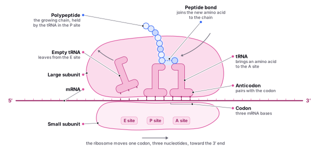
Find it in the figure
Coming soon for figure 9.1
Section 9.5
Answering Questions on Complementary DNA strands
When answering questions related to the concept of complementary DNA strands, base pairing, and RNA transcription, it is crucial to have a solid understanding of the basic principles. Here is a step-by-step guide to help you approach exam questions on these topics:
Review the key concepts: DNA is double-stranded and composed of complementary strands.
- Base pairing rules: Adenine (A) pairs with Thymine (T), and Cytosine (C) pairs with Guanine (G) in DNA. In RNA, Thymine (T) is replaced by Uracil (U).
- The coding strand contains the genetic information for protein synthesis, while the template strand is used to create a complementary RNA copy.
Determine the proportion of bases:
- Remember Chargaff's rules: The amount of adenine (A) equals the amount of thymine (T), and the amount of cytosine (C) equals the amount of guanine (G) in a DNA molecule.
- If the question provides the proportion or percentage of some bases, you can calculate the proportion of the remaining bases using Chargaff's rules.
Identify the coding and template strands:
- The coding strand has the same sequence as the resulting mRNA, with the exception that thymine (T) is replaced by uracil (U) in RNA.
- The template strand is complementary to both the coding strand and the resulting mRNA.
Calculate complementary sequences:
- If you are given a DNA sequence and asked to determine the complementary sequence, use the base pairing rules: A pairs with T, and C pairs with G.
- If you are given a template DNA strand and asked to determine the resulting mRNA sequence, use the base pairing rules, but remember to replace T with U in RNA: A pairs with U, and C pairs with G.
Now, let's apply this guide to some example questions:
Example 1: If 30 percent of the bases in a DNA molecule are adenine (A), what is the percentage of the other bases?
- According to Chargaff's rules, the percentage of thymine (T) will also be 30 percent (since A pairs with T).
- The remaining percentage (100 - 30 - 30 = 40 percent) is split equally between cytosine (C) and guanine (G), so each will be 20 percent.
Example 2: Given the template DNA strand 3'-TACGAT-5', what is the sequence of the coding strand and the resulting mRNA?
- The coding strand (5'-3') is complementary to the template strand: 5'-ATGCTA-3'.
- The resulting mRNA has the same sequence as the coding strand, with T replaced by U: 5'-AUGCUA-3'.