


Chapter 7IMAT Biology
The Cell Reproduction Cycle
Cell division is the process by which cells replicate and produce new cells. It is an essential process for the growth and repair of tissues and the continuation of life. Without cell division, organisms would not be able to grow, repair damaged tissues, or reproduce.
Cell division occurs in two main types: and meiosis. Mitosis is the process by which a single cell divides into two identical daughter cells, each with the same number of as the parent cell. Meiosis, on the other hand, is the process by which cells divide into four non-identical daughter cells with half the number of chromosomes as the parent cell.
During mitosis, the chromosomes in the parent cell are replicated and then divided equally into the two daughter cells. This ensures that each daughter cell has the same genetic information as the parent cell. Mitosis is essential for the growth and repair of tissues and the continuation of life.
Meiosis, is important for sexual reproduction. It occurs in the formation of eggs and sperm and produces four non-identical daughter cells with half the number of chromosomes as the parent cell. This ensures that when an egg and sperm combine during fertilization, the resulting offspring will have the appropriate number of chromosomes. But before Mitosis and Meiosis, the cell must undergo through the which allows it to prepare to the M phase (Meiosis or Mitosis)
Section 7.1
The Cell Cycle
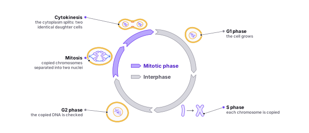
The cell cycle is the series of events that occurs in a cell as it grows and prepares to divide into daughter cells. It consists of two main phases: and cell division.
Interphase, which accounts for the majority of the cell cycle, is a period of intense activity where the cell prepares for division. During this time, the cell grows, duplicates its DNA, and synthesizes the proteins and other cellular components necessary for cell division. Interphase is divided into three main sub-phases:
: The G1 phase is particularly significant because it is the first phase of interphase, during which the cell grows and synthesizes the proteins and other cellular components needed for DNA replication and cell division. The cell also checks for DNA damage and any other abnormalities during this phase, ensuring that the DNA is suitable for replication.
: The S phase is critical because it is when DNA replication occurs. DNA replication is an essential process, as each daughter cell must receive a complete set of genetic material to function correctly. Any errors in DNA replication can lead to mutations, which can have severe consequences, including the development of cancer.
: The G2 phase is important for checking that the cell has replicated its DNA correctly and that there are no other errors in the genetic material. If any abnormalities are detected during the G2 phase, the cell will not enter the final phase of the cell cycle, known as cell division, until the errors have been corrected.
The second phase of the cell cycle is cell division, which is divided into two main types: mitosis and meiosis.
: Some cells may enter a state called G0 phase, where they are temporarily quiescent and do not actively divide. Cells in G0 phase may still perform their normal functions, but they are not actively preparing to divide. Examples of cells that may enter G0 phase include differentiated cells, such as some nerve cells and muscle cells, which do not divide after they have matured.
Mitosis: During mitosis, the cell's replicated chromosomes are evenly divided into two daughter cells, resulting in two identical daughter cells with the same number of chromosomes as the parent cell.
Meiosis: During meiosis, the cell's replicated chromosomes are divided into four non-identical daughter cells with half the number of chromosomes as the parent cell. This process is vital for sexual reproduction, as it ensures that the resulting offspring will have the appropriate number of chromosomes.
Find it in the figure
Coming soon for figure 7.1
The S phase
The S phase, or the synthesis phase, is the second phase of the interphase in the cell cycle. It is characterized by the replication of the cell's DNA, meaning a copy of each chromosome is made. This is an essential step in preparation for cell division, as it ensures that each daughter cell will have the same genetic information as the parent cell.
During the S phase, the number of DNA molecules in the cell increases from n to 2n. This is because each chromosome is replicated, resulting in two copies of each chromosome. An analogy to understand this process is to think of a book. Just as a book contains information that can be copied onto a second book, the DNA in a cell contains genetic information that can be copied onto the second set of chromosomes.
As an example, let's consider the fruit fly Drosophila melanogaster. The fruit fly has 4 pairs of chromosomes, for a total of 8 chromosomes. During the S phase, each of these 8 chromosomes is replicated, resulting in a total of 16 chromatids in the cell. These 16 chromatids are then evenly divided into two daughter cells during the process of mitosis, resulting in two daughter cells with 8 chromosomes each.
During the S phase, or synthesis phase, of the cell cycle, the cell's DNA is replicated. This process involves the synthesis of a new complementary strand of DNA for each existing strand of DNA, resulting in the doubling of the number of DNA molecules in the cell.
The significance of the S phase lies in its essential role in preserving the integrity of genetic information during cell division. By accurately replicating the cell's DNA, the S phase ensures that each daughter cell receives the same genetic information as the parent cell. This process is crucial for maintaining the normal functioning of cells, tissues, and organs, as well as ensuring the continuity of genetic information across generations.
Errors during the S phase can lead to various problems and medically relevant conditions, including:
- Mutations: Errors in DNA replication can introduce mutations, which are changes in the DNA sequence. These mutations can be harmless, beneficial, or detrimental depending on their nature and location in the genome. Some mutations can lead to genetic disorders, increase the risk of developing diseases, or cause cancer.
- Chromosomal abnormalities: Errors during the S phase can result in abnormal chromosome numbers or structures. These abnormalities can lead to conditions such as Down syndrome, Turner syndrome, or Klinefelter syndrome, which are caused by an extra or missing chromosome.
- Replication stress: Incomplete or stalled replication can cause replication stress, which can lead to DNA damage and genomic instability. This instability can contribute to the development of various diseases, including cancer.
- Cell cycle arrest and cell death: Errors during the S phase can activate cell cycle checkpoints, which halt cell division to allow time for repair. If the damage is too severe, the cell may undergo programmed cell death, or apoptosis, to prevent the propagation of faulty genetic information.
- Cancer: Many types of cancer are associated with errors in DNA replication during the S phase. These errors can lead to genomic instability, uncontrolled cell division, and the formation of tumors.
In summary, the S phase is essential for the accurate replication of genetic information, which is crucial for maintaining the normal functioning of cells and the continuity of genetic information. Errors during the S phase can have significant consequences, leading to various medical conditions and diseases, including cancer.
DNA Replication (In Detail)
DNA replication is the process by which cells create a copy of their genetic material, DNA, in preparation for cell division. It is a crucial process that ensures the accurate transmission of genetic information from one generation of cells to the next. In this section, we will delve into the details of DNA replication, including the enzymes involved, the direction of replication, and the role of Okazaki fragments.
Before we begin, it is important to understand the structure of DNA. DNA, or deoxyribonucleic acid, is a double-stranded molecule made up of two strands of nucleotides that are held together by hydrogen bonds. Each nucleotide consists of a sugar molecule called deoxyribose, a phosphate group, and one of four nitrogenous bases: adenine (A), cytosine (C), guanine (G), or thymine (T). The order of these bases, or the sequence of the DNA, is what carries the genetic information.
Now, let's talk about the process of DNA replication. DNA replication occurs in the S phase of the cell cycle, which is the period between cell division. It starts with the unwinding of the double helix structure of the DNA molecule by an enzyme called helicase. This creates a replication fork, where the two strands of the DNA molecule separate and expose the bases.
Next, an enzyme called primase adds short RNA primers to the template strands of DNA. These primers serve as a starting point for the synthesis of new strands of DNA.
Once the primers are in place, another enzyme called polymerase starts synthesizing the new strands of DNA. There are several different types of polymerases, but the one primarily responsible for DNA replication is called DNA polymerase III. DNA polymerase III adds nucleotides to the template strands of DNA by matching the correct base to the template strand using the base-pairing rule: A with T, and C with G. This process is called semi-conservative replication because one of the original strands is conserved and used as a template for the synthesis of the new strand.
As DNA polymerase III moves along the template strand, it synthesizes the new strand in the 5' to 3' direction, meaning that it starts at the 5' end and moves towards the 3' end. This is important because it ensures that the new strands are synthesized in the correct order and that the genetic information is accurately transmitted.
However, there is a problem with this process. DNA polymerase III can only add nucleotides in the 5' to 3' direction, which means that it cannot synthesize the new strand continuously. Instead, it has to synthesize short stretches of DNA called Okazaki fragments. These fragments are named after the scientist who discovered them, Reiji Okazaki.
Once an Okazaki fragment is synthesized, an enzyme called ligase seals the gap between the fragments by creating a covalent bond between the sugar and phosphate groups. This creates a continuous strand of DNA.
During DNA replication, the leading strand, which is the strand being synthesized in the same direction as the replication fork, can be synthesized continuously by DNA polymerase III. However, the lagging strand, which is the strand being synthesized in the opposite direction of the replication fork, is synthesized in a discontinuous manner, with Okazaki fragments being synthesized and then sealed together by ligase.
In summary, DNA replication is a complex process that involves several enzymes, including helicase, primase, DNA polymerase III, and ligase. It is a semi-conservative process that involves the synthesis of new strands of DNA using the template strands as a guide. The direction of replication is from 5' to 3' and is important for the accurate transmission of genetic information. Okazaki fragments play a crucial role in the synthesis of the lagging strand, which is synthesized in a discontinuous manner.
It is worth noting that the process of DNA replication is not always perfect and mistakes, or errors, can occur. These errors, called mutations, can alter the sequence of the DNA and potentially have consequences for the organism. To ensure the accuracy of DNA replication, cells have a number of mechanisms in place to detect and repair errors.
One such mechanism is proofreading, which is carried out by DNA polymerase III. DNA polymerase III has the ability to recognize and remove mismatched bases before they are incorporated into the new strand of DNA. Another mechanism is mismatch repair, which involves the identification and correction of mismatched bases after they have been incorporated into the new strand.
In addition to these mechanisms, cells also have a number of enzymes called exonucleases that can remove nucleotides from the ends of DNA strands. These enzymes can be used to remove any excess nucleotides that may have been added during replication.
Overall, DNA replication is a vital process that ensures the accurate transmission of genetic information from one generation of cells to the next. Understanding the details of this process, including the enzymes involved and the role of Okazaki fragments, is important for a number of fields, including molecular biology and medicine.
The Important Aspects of DNA Replication
DNA replication is a complex process that involves many different components and enzymes working together to ensure the accurate duplication of genetic information. Here are some of the most important aspects of DNA replication:
- Helicase: This enzyme is responsible for unwinding the double-stranded DNA helix to form a replication fork, which is the site where replication occurs. Helicase does this by breaking the hydrogen bonds that hold the two strands of DNA together. It moves along the DNA strand in a 5' to 3' direction, separating the strands and creating a single-stranded template for replication. Helicase is essential for DNA replication to occur, as the double-stranded DNA helix must be unwound before the polymerases can begin to synthesize new strands.
- Single-strand binding proteins: These proteins bind to and stabilize single-stranded DNA, preventing it from forming secondary structures that would interfere with replication. They coat the single-stranded template, preventing it from re-forming into a double-stranded helix. Without these proteins, the single-stranded template would quickly become re-paired with its complementary strand, halting the replication process.
- Primase: This enzyme synthesizes a short RNA primer at the replication fork, providing a 3'-OH group to which DNA polymerase can add nucleotides. Primase is essential for DNA replication because DNA polymerase can only add nucleotides to an existing 3'-OH group. Without the primer provided by primase, DNA polymerase would not be able to start synthesizing the new DNA strands.
- DNA polymerase: This enzyme is responsible for extending the RNA primer by adding nucleotides to the 3'-OH group. There are multiple DNA polymerases involved in DNA replication, each with a different function. DNA polymerase III is the main polymerase responsible for elongating the new DNA strands, while DNA polymerase I is involved in removing the RNA primers and replacing them with DNA. DNA polymerase also has a proofreading function, which allows it to correct errors in base pairing that may occur during replication.
- DNA ligase: This enzyme joins the Okazaki fragments on the lagging strand by catalyzing the formation of a phosphodiester bond between adjacent nucleotides. DNA ligase is essential for DNA replication because the lagging strand is synthesized in short fragments that must be joined together to form a continuous strand.
- Topoisomerase: This enzyme relaxes the supercoiling ahead of the replication fork, preventing the DNA strands from becoming too tightly wound. Supercoiling occurs when the DNA helix twists upon itself as it unwinds, and if left unchecked, it can cause breaks in the DNA strand. Topoisomerase relieves this tension by breaking the DNA strand, allowing it to unwind, and then re-joining the strand.
- Proofreading: DNA polymerase proofreads newly synthesized DNA, correcting errors in base pairing. This function ensures that the new DNA strands are as accurate as possible, minimizing the risk of mutations and errors that can lead to disease.
- Telomerase: This enzyme extends the ends of linear chromosomes by adding repetitive DNA sequences called . Telomeres are essential for protecting the ends of chromosomes from degradation and fusion with other chromosomes. Without telomeres, the ends of chromosomes would gradually erode with each cell division, leading to genomic instability and cell death.
- Replication origin: This is the specific sequence of DNA where replication begins. Replication origin sequences are recognized by proteins that initiate replication by recruiting helicase and other replication enzymes to the site. The location and number of replication origins varies between organisms and cell types, depending on the size and complexity of their genomes.
Understanding these different aspects of DNA replication is essential for understanding how genetic information is transmitted from one generation to the next, and how mutations and errors can occur during replication.
Section 7.2
Mitosis
Mitosis is a vital cellular process that results in a single cell dividing into two genetically identical daughter cells, each possessing the same number of chromosomes as the parent cell. This process is crucial for the growth, repair, and maintenance of tissues, as well as the perpetuation of life.
Mitosis can be subdivided into four primary stages, followed by , the actual division of the cytoplasm:
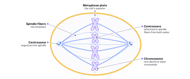
- : Prophase marks the beginning of mitosis. During this stage, the in the cell's nucleus condenses into visible chromosomes, and the nucleolus disappears. Each chromosome consists of two identical joined at the . The centrosomes, which are responsible for organizing the mitotic spindle, begin to migrate to opposite poles of the cell. The mitotic spindle, composed of microtubules, starts to form between the centrosomes. Additionally, the nuclear envelope starts to break down, allowing the spindle fibers to access the chromosomes.
- : In metaphase, the chromosomes align along the cell's equator, forming the metaphase plate. Each chromosome's centromere becomes attached to the spindle fibers, which extend from the centrosomes at the cell's poles. This connection ensures that each daughter cell will receive one copy of each chromosome during cell division.
- : Anaphase is characterized by the separation of sister chromatids. The centromere of each chromosome splits, and the sister chromatids are pulled to opposite poles of the cell by the mitotic spindle fibers. This movement ensures that each daughter cell will have an identical set of chromosomes.
- : During telophase, the cell starts to divide into two daughter cells. A new nuclear envelope forms around each set of chromosomes, and the nucleolus reappears within each nucleus. The chromosomes begin to decondense back into chromatin, and the mitotic spindle disassembles.
- Cytokinesis: Cytokinesis is the final stage of cell division, during which the cytoplasm and cellular organelles are divided between the two emerging daughter cells. In animal cells, a contractile ring composed of actin and myosin filaments constricts at the cell's equator, creating a cleavage furrow. This furrow deepens until the cell is pinched into two separate cells. In plant cells, a cell plate forms between the two daughter cells, eventually developing into a new cell wall, which separates the two cells.
Find it in the figure
Coming soon for figure 7.2
In summary, mitosis is a critical process that ensures the accurate distribution of genetic material to two daughter cells. It consists of four main stages—prophase, metaphase, anaphase, and telophase—followed by cytokinesis. This process plays a pivotal role in the growth, repair, and continuation of life across various organisms.
Section 7.3
Meiosis
Meiosis is a specialized cell division process that results in four non-identical daughter cells, each with half the number of chromosomes as the parent cell. This process is crucial for sexual reproduction, as it ensures that offspring will have the appropriate number of chromosomes. Meiosis consists of two main stages: meiosis I and meiosis II, each with its respective substages.
Meiosis I: Meiosis I comprises the following substages:
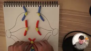
- Prophase I: During prophase I, chromosomes condense and become visible. The nuclear envelope begins to break down, and the spindle fibers start to form. Homologous chromosomes (one from each parent) come together in a process called synapsis, forming a structure called a bivalent or tetrad. At this stage, genetic material may be exchanged between non-sister chromatids in a process called crossing over, which generates genetic diversity in the resulting gametes.
- Metaphase I: In metaphase I, the homologous chromosome pairs align at the cell's equator, forming two columns, unlike the single column seen in mitosis. This arrangement is due to the prior synapsis, where chromosomes paired up with their homologous partners. Spindle fibers attach to the centromeres of each homologous chromosome pair.
- Anaphase I: During anaphase I, the spindle fibers pull the homologous chromosome pairs to opposite poles of the cell. Unlike mitosis, the sister chromatids remain attached at their centromeres and do not separate.
- Telophase I: In telophase I, the chromosomes reach the poles, and the cell begins to divide, resulting in two daughter cells. Each daughter cell has half the number of chromosomes as the parent cell, but the chromosomes still consist of two chromatids.
- Cytokinesis I: Cytokinesis I follows telophase I, resulting in the separation of the two daughter cells.
Meiosis II: Meiosis II is similar to mitosis and includes the following substages:
- Prophase II: During prophase II, the nuclear envelope dissolves again in the two daughter cells, and the spindle fibers begin to form.
- Metaphase II: In metaphase II, the chromosomes align at the equator of each daughter cell, with spindle fibers attaching to the centromeres.
- Anaphase II: During anaphase II, the centromeres of each chromosome finally split, and the sister chromatids are pulled to opposite poles of the cell by the spindle fibers.
- Telophase II: In telophase II, the chromatids reach the poles, and the nuclear envelope starts to reform around the separated chromatids, which are now considered individual chromosomes.
- Cytokinesis II: Cytokinesis II follows telophase II, dividing each daughter cell from meiosis I into two new cells, resulting in a total of four non-identical daughter cells, each with half the number of chromosomes as the parent cell.
In summary, meiosis is a critical process for sexual reproduction, generating non-identical daughter cells with half the number of chromosomes as the parent cell. This process comprises two main stages, meiosis I and meiosis II, each with its own substages. Crossing over during prophase I contributes to genetic diversity among the resulting gametes, which is essential for the evolution and adaptation of species.
Prophase I
Prophase 1 is the first stage of meiosis, a type of cell division that produces gametes, or sex cells, such as sperm and eggs. In meiosis, a single cell divides into four daughter cells, each of which contains half the number of chromosomes as the parent cell. This reduction in chromosome number is important for sexual reproduction, as it allows for the exchange of genetic material between the parents and the production of genetically diverse offspring.
During prophase 1, the chromosomes in the nucleus of the cell become visible and begin to condense. This is important because it allows for the precise separation of the chromosomes during the subsequent stages of meiosis. The chromosomes are made up of DNA, which is tightly packed into a compact structure called chromatin. During prophase 1, the chromatin condenses and becomes visible under the microscope as discrete threads called chromosomes.
As prophase 1 progresses, the nucleolus disappears and the nuclear envelope breaks down. The centrosomes, which are responsible for organizing the mitotic spindle, also begin to move to opposite poles of the cell. The mitotic spindle is a structure that is composed of microtubules and is responsible for separating the chromosomes during cell division.
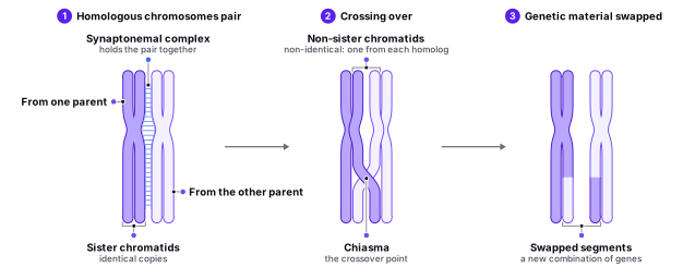
One of the key events that occurs during prophase 1 is crossing over, also known as recombination. This is the exchange of genetic material between homologous chromosomes, which are pairs of chromosomes that contain the same genes in the same order. Crossing over occurs during prophase 1 and allows for the production of genetically diverse offspring by shuffling the genetic material of the parents.
The exchange of genetic material between homologous chromosomes occurs at specific points called chiasmata, which are visible as small, X-shaped structures under the microscope. Chiasmata form when the homologous chromosomes pair up and exchange genetic material. This process is facilitated by a protein structure called the synaptonemal complex, which holds the chromosomes in close proximity to each other.
During crossing over, the homologous chromosomes pair up and the synaptonemal complex forms between them. The chromosomes then exchange genetic material at the chiasmata, creating a new combination of genes on each chromosome. This process can occur at multiple points along the length of the chromosomes, resulting in the shuffling of genetic material.
After crossing over is complete, the homologous chromosomes begin to separate and move towards the equator of the cell. This is the beginning of metaphase 1, the next stage of meiosis.
In summary, prophase 1 is the first stage of meiosis and is characterized by the condensation of the chromosomes, the breakdown of the nuclear envelope, and the movement of the centrosomes to opposite poles of the cell. It is also the stage during which crossing over, or recombination, occurs, allowing for the exchange of genetic material between homologous chromosomes. The exchange of genetic material occurs at chiasmata, which are facilitated by the synaptonemal complex. Understanding the process of prophase 1 is important for understanding the mechanisms of sexual reproduction and the production of genetically diverse offspring.
Find it in the figure
Coming soon for figure 7.3
Prophase I Steps
Leptotene, zygotene, pachytene, diplotene, and diakinesis are stages of prophase 1 in meiosis, a type of cell division that produces gametes, or sex cells, such as sperm and eggs. During meiosis, a single cell divides into four daughter cells, each of which contains half the number of chromosomes as the parent cell. This reduction in chromosome number is important for sexual reproduction, as it allows for the exchange of genetic material between the parents and the production of genetically diverse offspring.
Leptotene is the first stage of prophase 1 and is characterized by the appearance of thin, elongated chromosomes under the microscope. The chromosomes are made up of DNA, which is tightly packed into a compact structure called chromatin. During leptotene, the chromatin begins to condense and becomes visible as discrete threads called chromosomes.
Zygotene is the next stage of prophase 1 and is characterized by the formation of the synaptonemal complex, a protein structure that holds homologous chromosomes, or pairs of chromosomes that contain the same genes in the same order, in close proximity to each other. The synaptonemal complex is visible under the microscope as a series of parallel lines between the homologous chromosomes.
Pachytene is the third stage of prophase 1 and is characterized by the complete formation of the synaptonemal complex and the beginning of crossing over, also known as recombination. During crossing over, the homologous chromosomes exchange genetic material at specific points called chiasmata, which are visible as small, X-shaped structures under the microscope. This process shuffles the genetic material of the parents and allows for the production of genetically diverse offspring.
Diplotene is the fourth stage of prophase 1 and is characterized by the separation of the homologous chromosomes. The synaptonemal complex begins to break down and the chromosomes start to move apart, held together only at the chiasmata.
Diakinesis is the final stage of prophase 1 and is characterized by the further separation of the homologous chromosomes and the formation of the mitotic spindle. The mitotic spindle is a structure that is composed of microtubules and is responsible for separating the chromosomes during cell division.
In summary, leptotene, zygotene, pachytene, diplotene, and diakinesis are stages of prophase 1 in meiosis. They are characterized by the condensation of the chromosomes, the formation and breakdown of the synaptonemal complex, and the exchange of genetic material between homologous chromosomes through crossing over. Understanding these stages is important for understanding the mechanisms of sexual reproduction and the production of genetically diverse offspring.
Section 7.4
The Main Differences Between Mitosis and Meiosis
Mitosis and meiosis are both essential cell division processes, but they serve different purposes and have distinct outcomes. Thus, now that you are aware of the different processes that are involved in each, it will be useful to consolidate all of this information by appreciating the differences among those two. Here is a detailed comparison of their main differences to better understand their respective significance:
Purpose:
- Mitosis is responsible for growth, repair, and maintenance of tissues in an organism. It is also involved in asexual reproduction, where a single parent cell divides to produce genetically identical offspring.
- Meiosis, on the other hand, is crucial for sexual reproduction, as it produces gametes (sperm and egg cells) with half the number of chromosomes as the parent cell. This ensures that the resulting offspring will have the appropriate number of chromosomes when gametes fuse during fertilization.
Number of divisions:
- Mitosis involves a single cell division, resulting in two genetically identical daughter cells.
- Meiosis comprises two cell divisions (meiosis I and meiosis II), resulting in four genetically unique daughter cells.
Chromosome number in daughter cells:
- In mitosis, the daughter cells have the same number of chromosomes as the parent cell (diploid).
- In meiosis, the daughter cells have half the number of chromosomes as the parent cell (haploid).
Genetic variation:
- Mitosis produces genetically identical daughter cells, with no variation in genetic material.
- Meiosis generates genetic diversity through two processes: crossing over during prophase I, which involves the exchange of genetic material between homologous chromosomes, and independent assortment during metaphase I, where the homologous chromosomes align randomly at the cell's equator.
Type of cells involved:
- Mitosis occurs in somatic (non-reproductive) cells.
- Meiosis takes place in germ cells, which are specialized cells that give rise to gametes (sperm and egg cells).
Chromosome alignment:
- In mitosis, individual chromosomes line up along the cell's equator during metaphase, forming a single column.
- In meiosis I, homologous chromosomes pair up and align along the cell's equator during metaphase I, forming two columns.
Sister chromatid separation:
- In mitosis, sister chromatids separate and are pulled to opposite poles during anaphase.
- In meiosis, sister chromatids do not separate during anaphase I; instead, homologous chromosomes are pulled apart. Sister chromatids separate during anaphase II of meiosis II.
By understanding these key differences between mitosis and meiosis, students can better appreciate their respective roles in growth, repair, and reproduction, as well as the generation of genetic diversity in sexually reproducing organisms.