







Chapter 3IMAT Biology
Cell Biology
As you may already know, cells are the fundamental unit of life, and they come in a wide variety of shapes and sizes, each with its own unique functions and roles. In this chapter, we will embark on a journey through the complex and intricate world of cells, exploring their structure, function, and role in the overall functioning of living organisms.
As you delve into this chapter, you will learn about the different types of cells found in the natural world, including prokaryotic cells and eukaryotic cells. You will also discover the intricate structure of cells, including their various organelles and their functions, such as the mitochondria, which produce energy for the cell, and the endoplasmic reticulum and Golgi apparatus, which are involved in protein synthesis and transport.
In addition to learning about the structure and function of cells, we will also delve into the process of cell division, which is essential for the growth and repair of living organisms. You will learn about and meiosis, the two types of cell division, and how they allow cells to replicate and pass on genetic information to the next generation.
But cells are not just isolated units; they are also highly interactive and communicate with each other in a variety of ways. In this chapter, we will discuss the various mechanisms by which cells communicate, including through chemical signaling, cell-cell adhesion, and extracellular matrix.
“Cell Biology” is the study of cells, which are the basic unit of life. All living things are made up of cells, and they perform all of the functions necessary for life, such as growth, metabolism, and reproduction.
Cells come in many different shapes and sizes, and they can be found in every living organism on Earth, from tiny bacteria to complex animals like humans. Cells are the building blocks of life, and they are responsible for all of the functions necessary for an organism to survive.
One of the most exciting things about cells is that they have their own internal structures and organelles, which are specialized parts that perform specific functions. For example, the mitochondria are the “powerhouses” of the cell, responsible for generating energy in the form of a molecule called “”. The nucleus is the “brain” of the cell, containing the genetic material that controls all of the cell's activities.
Prokaryotic cells, are usually smaller and simpler in structure than eukaryotic cells. They are found in single-celled organisms, such as bacteria. Prokaryotic cells do not have a nucleus or other membrane-bound organelles. Instead, their genetic material is found in a single loop of DNA floating in the cytoplasm. Bacteria also have loops of genetic material called “Plasmids”, they have a similar role, and allow the bacteria to gain different characteristics rather quickly, such as antibiotic resistance, by transferring the plasmids to the current, or the next generation of bacteria.
Eukaryotic cells are found in more complex organisms, such as plants and animals. They are larger and more complex than prokaryotic cells and have a defined nucleus and membrane-bound organelles.
Membrane-bound organelles are subcellular structures that are enclosed within their own lipid bilayer membrane. They are found in eukaryotic cells, which are cells that have a true nucleus and other membrane-bound organelles. Membrane-bound organelles are specialized structures that perform specific functions within the cell.
The lipid bilayer membrane that encloses these organelles serves several important functions. It acts as a barrier, separating the contents of the organelle from the rest of the cell. It also helps maintain the organelle's structural integrity and regulates the flow of materials in and out of it.
Some other important organelles in eukaryotic cells include the endoplasmic reticulum, which is involved in the synthesis and transport of proteins and lipids, and the Golgi apparatus, which is involved in the modification, sorting, and delivery of proteins and lipids to their final destination. Don't worry! We will soon learn about all of these organelles one by one!
The cell membrane separates the inside of the cell from the outside environment and helps to maintain the cell's shape. The cell membrane is mostly made up of lipids and proteins, and it is selectively permeable, allowing certain substances to pass through while blocking others.
In addition to the organelles mentioned earlier, a few other important structures are found in cells that are worth discussing.
One of these important structures is the cytoskeleton, a network of protein fibers that gives the cell its shape and helps it maintain its structure. The cytoskeleton comprises three types of protein fibers: microtubules, microfilaments, and intermediate filaments. These fibers can be found throughout the cell. They play a vital role in many of the cell's activities, including movement, division, and the transport of materials within the cell.
Some protist, plant, and fungi cells have a structure called the cell wall, a different structure than the cell membrane, which serves as a protective layer. The cell wall helps to give the cell its shape and provides extra support and protection.
Section 3.1
Membrane-bound Organelles
Membrane-bound organelles are specialized structures within a cell enclosed by a membrane. These organelles perform specific functions necessary for the cell to carry out its various activities. On the IMAT exam, there's a list of essential organelles worth learning. Let's review all of the organelles we will cover in-depth later on:
- The nucleus: The nucleus is the central organelle of the cell and is responsible for storing and processing genetic information. It is surrounded by a double lipid bilayer membrane called the nuclear envelope, which separates the contents of the nucleus from the rest of the cell.
- The endoplasmic reticulum (ER): The endoplasmic reticulum (ER) is a network of flattened sacs and tubes that is involved in the synthesis, modification, and transport of proteins and lipids. The ER is divided into two main types: the smooth endoplasmic reticulum (SER) and the rough endoplasmic reticulum (RER). The SER is involved in the synthesis of lipids, while the RER is involved in the synthesis of proteins.
- The Golgi apparatus: The Golgi apparatus is a stack of flattened membrane-bound sacs that is involved in the sorting, modification, and transport of proteins and lipids. It receives newly synthesized molecules from the endoplasmic reticulum and sorts them into vesicles for transport to their final destination.
- The mitochondria:The mitochondria are the cell's powerhouses, responsible for producing the energy that the cell needs to carry out its various functions. They are surrounded by a double membrane and contain their own DNA and ribosomes.
- The lysosomes: Lysosomes are small, membrane-bound organelles that contain hydrolytic enzymes that are responsible for breaking down waste materials and cellular debris. They help to maintain the cell's internal environment by removing unnecessary or damaged materials.
- The peroxisomes: Peroxisomes are small, membrane-bound organelles that contain enzymes that are involved in the breakdown of fatty acids and the detoxification of harmful substances. They are similar to lysosomes in function, but they do not contain the same types of hydrolytic enzymes.
- The vacuoles: Vacuoles are large, membrane-bound organelles that are involved in the storage of various substances, including water, salts, and pigments. Plant cells typically have one or more large central vacuoles that are used to store water and other substances, while animal cells have smaller vacuoles that are involved in the storage of specific substances.
Section 3.2
Prokaryotic Cells
Prokaryotic cells are the simplest and most ancient type of cells. Prokaryotes are single-celled organisms that lack a nucleus and other membrane-bound organelles. They are found in a variety of environments, including soil, water, and the human body. Examples of prokaryotes include bacteria and archaea.
Despite their simple structure, prokaryotes play an important role in the biological ecosystem. They are involved in nutrient cycling and play a crucial role in the decomposition of organic matter. They are also important in the production of certain foods such as cheese, yogurt, and vinegar. Some types of bacteria can cause disease, while others are beneficial to human health, such as those found in the human gut microbiome.
They are characterized by the absence of a true nucleus and membrane-bound organelles. Instead, their genetic material is found in a single, circular that is located in the cytoplasm, in an area called “nucleoid”. Prokaryotes are usually much smaller than eukaryotic cells as most common bacteria are about 1 to 2 microns in diameter and 1 to 4 microns long, on average.
Prokaryotes are classified into two main groups: Bacteria and Archaea. Bacteria are the most well-known type of prokaryotes and are found in almost every environment on Earth. They are important for a variety of ecological functions, including the decomposition of organic matter, the nitrogen cycle, and the production of various substances that are used by other organisms. Archaea are a group of prokaryotes that are adapted to extreme environments, such as hot springs, salt flats, and the deep sea.
Prokaryotes are generally classified based on their shape, which can be rod-shaped (bacilli), spherical (cocci), or spiral (spirilla). They can also be classified based on their method of obtaining energy, which can be through photosynthesis (phototrophs) or through the breakdown of organic matter (heterotrophs). Prokaryotes are adapted for survival in a variety of environments and have a range of mechanisms for obtaining nutrients, such as the ability to take up nutrients from the environment or to produce their own nutrients through photosynthesis. They also have a range of mechanisms for defending against predators and environmental stresses, such as the production of antibiotics or the ability to form endospores.
Endospores allows the bacterium to produce a dormant and highly resistant cell to protect the cell's genetic material in times of extreme stress, such as high UV radiation and temperatures.
Prokaryotes are important in many different fields, including medicine, agriculture, and environmental science. They are also important models for understanding basic cellular processes, as they are much simpler and easier to study than eukaryotic cells.
Prokaryotic Structure
Prokaryotic cells are the simplest and most ancient type of cells. They are characterized by the absence of a true nucleus and membrane-bound organelles. Instead, their genetic material is found in a single, circular chromosome that is located in the cytoplasm.
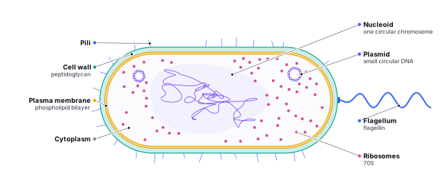
The basic structure of a prokaryotic cell consists of the following components:
- Cell wall:The cell wall is a rigid, protective layer that surrounds the cell and gives it its shape. It is made up of peptidoglycan, a complex carbohydrate-protein compound that is unique to prokaryotes.
- Plasma membrane: The plasma membrane is a thin, flexible barrier that surrounds the cell and separates the inside from the outside. It is made up of a phospholipid bilayer and is responsible for maintaining the cell's internal environment and regulating the movement of substances in and out of the cell.
One major difference is that the phospholipid bilayers of eukaryotic cells are typically thicker than those of bacterial cells. This is due to the presence of additional lipids and proteins within the bilayer of eukaryotic cells, which help to form specialized structures which are involved in various processes such as endocytosis, exocytosis, and signaling.
Another difference is that the phospholipid bilayers of eukaryotic cells are more complex and dynamic, with a greater variety of lipids and proteins present. In contrast, bacterial phospholipid bilayers are relatively simple, with only a few types of lipids and proteins present.
Finally, the phospholipid bilayers of eukaryotic cells are more tightly packed and oriented in a specific direction, while those of bacterial cells are more disordered and fluid. This difference in packing and orientation is due to the presence of proteins and lipids with specific shapes and properties within the eukaryotic bilayer.
- Cytoplasm: The cytoplasm is the gel-like substance that fills the cell and surrounds the genetic material. It contains all of the cell's metabolic machinery, including enzymes, ribosomes, and other cellular structures.
There are several differences between the cytoplasm of bacteria and eukaryotes crucial to remember to the IMAT exam.
One major difference is the presence of a cytoskeleton in eukaryotes. The cytoskeleton is a network of protein fibers that provides structural support and helps to maintain the shape of eukaryotic cells. It is composed of three types of fibers: microtubules, microfilaments, and intermediate filaments. Bacteria do not have the cytoskeleton, therefore do not have microtubules, microfilaments, or intermediate filaments. Research recently showed that some bacteria have its own “cytoskeleton”, which comprised of different proteins with similar function.
Another difference is the presence of membrane-bound organelles in eukaryotes. Membrane-bound organelles are specialized subcellular structures that perform specific functions within the cell. Examples of organelles found in eukaryotes include the nucleus, mitochondria, Golgi apparatus, and endoplasmic reticulum. Bacteria do not have membrane-bound organelles, but they may have non-membrane bounds structures, such as ribosomes and nucleoids.
A third difference is the presence of a nuclear envelope in eukaryotes. The nuclear envelope is a double-membraned structure that surrounds the nucleus and separates it from the cytoplasm. It is involved in controlling the movement of materials in and out of the nucleus. Bacteria do not have a nuclear envelope, but they may have a nucleoid, which is a region within the cell that contains the genetic material.
Overall, the cytoplasm of eukaryotes is more complex and organized than that of bacteria, due to the presence of organelles, a cytoskeleton, and a nuclear envelope. These structures allow eukaryotes to perform a wider range of functions and to have more specialized and compartmentalized cell biology.
- Genetic material:The genetic material of a prokaryotic cell is found in a single, circular chromosome that is located in the cytoplasm. It contains all of the genetic information needed for the cell to carry out its functions.
Plasmids are small, circular pieces of DNA that are found in some bacterial cells. They are separate from the bacterial chromosome and can replicate independently of it. Plasmids are usually present in small numbers in a cell, but under certain conditions, their population can increase.
Plasmids are often used as tools in molecular biology because they can be easily manipulated and introduced into bacterial cells. They can carry genetic material from one organism to another, which is why they are sometimes referred to as “vehicles” for genetic information. Plasmids can also be used to express proteins or to modify the properties of bacterial cells.
Plasmids are typically found in bacteria that live in environments with changing conditions, such as soil or the human gut. They allow bacteria to adapt to their environment by providing them with additional functions, such as the ability to break down certain compounds or to resist antibiotics.
- Bacterial Ribosomes: Ribosomes are small, spherical structures that are found in the cytoplasm and are involved in the synthesis of proteins.
- Flagella: Flagella are long, whip-like appendages that are used by some prokaryotes for movement. They are composed of a protein called flagellin and are attached to the cell wall.
- Pili: Pili are short, hair-like appendages that are found on the surface of some prokaryotes. They are used for a variety of purposes, including attachment to surfaces, transfer of genetic material, and communication with other cells.
Find it in the figure
Coming soon for figure 3.1
Bacterial Reproduction
Bacteria are prokaryotic cells that can reproduce through several methods. The most common method of bacterial reproduction is binary fission. In binary fission, a single cell divides into two identical daughter cells. The process of binary fission starts when the bacterial cell's genetic material replicates, or copies itself. Once the genetic material has been replicated, the cell begins to grow and elongate, and a new cell wall begins to form down the center of the cell, creating two separate compartments. The two copies of the genetic material are then separated and moved into the two compartments, and the cell eventually splits into two separate daughter cells.
Another method of bacterial reproduction is budding. Some bacteria are capable of budding, in which a new cell grows as an outgrowth from the parent cell and eventually detaches to form a separate cell. This is different from binary fission, where the cell splits into two identical daughter cells. In budding, the new cell is not identical to the parent cell, as it may have received different amounts of cytoplasm or genetic material.
Fragmentation is another method of bacterial reproduction. This occurs when a parent cell breaks into multiple fragments, each of which can develop into a new cell. Fragmentation can occur spontaneously, or it can be induced by environmental factors such as nutrient depletion or physical trauma.
Each of these methods of bacterial reproduction has its advantages and disadvantages. Binary fission is the most efficient method for producing large populations of bacteria quickly, while budding and fragmentation may allow bacteria to adapt to changing environmental conditions. Genetic recombination, on the other hand, allows bacteria to acquire new traits that may be advantageous in certain environments. Overall, the ability of bacteria to reproduce rapidly and adapt to changing conditions has made them important players in many ecological, industrial, and medical contexts.
Horizontal Gene Transfer
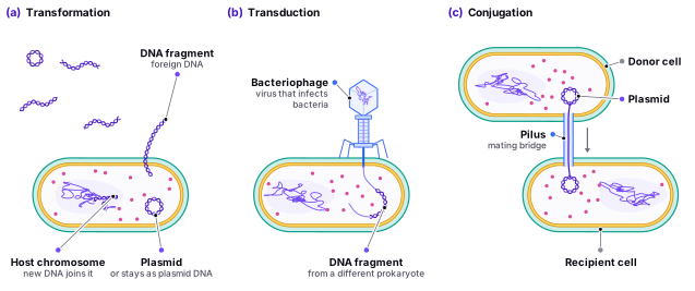
Horizontal gene transfer is the transfer of genetic material from one organism to another, without the involvement of reproduction. This process is particularly common in prokaryotes, which can exchange genetic material through several mechanisms, including transformation, transduction, and conjugation. These mechanisms are different from traditional reproduction, which involves the passing of genetic material from one generation to the next.
Transformation is the process by which bacteria take up foreign DNA from their surroundings and incorporate it into their own genome. This foreign DNA can come from other bacteria, viruses, or even the environment. Once incorporated into the bacterial genome, the new genetic material can be expressed and inherited by subsequent generations of bacteria. For example, Streptococcus pneumoniae bacteria can take up DNA fragments from dead bacterial cells in their environment, allowing them to acquire antibiotic resistance genes.
Transduction occurs when a virus transfers genetic material from one bacterial cell to another. In this process, a virus infects a bacterial cell and incorporates a fragment of the bacterial genome into its own DNA. When the virus infects another bacterial cell, it transfers the incorporated genetic material along with its own DNA. This can result in the transfer of genes between bacteria that are not closely related. For example, bacteriophages, viruses that infect bacteria, can transfer antibiotic resistance genes from one bacterium to another.
Conjugation is a more complex process in which bacterial cells transfer genetic material directly to each other through a specialized appendage called a pilus. In this process, a donor cell forms a pilus that attaches to a recipient cell, and a DNA molecule called a plasmid is transferred from the donor cell to the recipient cell. The plasmid can contain genes for antibiotic resistance, virulence factors, or other traits that are advantageous to the bacterial cell. For example, some strains of Escherichia coli can transfer a plasmid that contains genes for toxin production to other E. coli cells.
Transformation is a specific type of horizontal gene transfer in which a bacterial cell takes up and incorporates linear DNA molecules from the environment. These DNA molecules can come from other bacteria, viruses, or even eukaryotes. Transformation allows bacteria to acquire new genetic material and to adapt to new environments. For example, the bacterium Acinetobacter baylyi is known for its ability to take up DNA fragments from the environment and incorporate them into its genome, allowing it to adapt to a variety of environmental conditions.
In conclusion, horizontal gene transfer is an important mechanism that allows prokaryotes to exchange genetic information and to rapidly adapt to changing environments. Transformation, transduction, and conjugation are three different ways in which bacteria can transfer genetic material between cells. Transformation is a specific type of horizontal gene transfer that involves the uptake of linear DNA molecules from the environment. These mechanisms allow prokaryotes to acquire new genetic material and to evolve new traits, contributing to their diversity and adaptability.
Find it in the figure
Coming soon for figure 3.2
Section 3.3
Eukaryotic Cells
Have you ever wondered how complex organisms like plants, animals, and fungi are made up of so many different cells, each with its own specialized functions? The answer lies in the incredible world of eukaryotic cells. Eukaryotic cells are the building blocks of life as we know it, and they are responsible for the incredible diversity and complexity of the living world.
Eukaryotic cells are larger and more complex than their simpler, smaller counterparts, the prokaryotic cells. They have a true nucleus, which contains the cell's genetic material, or DNA, and is surrounded by a double membrane. This genetic material is organized into chromosomes, which carry the instructions that control the cell's functions and characteristics.
In addition to the nucleus, eukaryotic cells have a variety of other membrane-bound organelles, including the endoplasmic reticulum (ER), Golgi apparatus, mitochondria, and lysosomes. These organelles work together to carry out the many functions necessary for the cell to survive and thrive.
One of the most important features of eukaryotic cells is their ability to form tissues and organs, which work together to carry out the functions of the organism as a whole. For example, the cells that make up a plant's leaves work together to photosynthesize and produce energy for the plant, while the cells that make up a human's heart work together to pump blood throughout the body.
So, while eukaryotic cells may seem complex and mysterious, they are truly the building blocks of life. By studying these incredible cells, we can begin to understand the incredible diversity and complexity of the living world around us.
The Cytoplasm
The cytoplasm is a complex, gel-like substance that fills the cell and surrounds its organelles. It is composed of water, ions, and a variety of organic molecules, including enzymes, nucleic acids, and small organic molecules such as amino acids and sugars. The cytoplasm is a dynamic environment that is constantly changing and adapting to the needs of the cell.
One of the main functions of the cytoplasm is to provide a medium in which chemical reactions can occur. The cytoplasm contains a wide variety of enzymes that catalyze the chemical reactions necessary for the cell to function. These enzymes are involved in processes such as metabolism, energy production, and the synthesis of proteins, lipids, and other molecules. For example, the cytoplasm of a plant cell contains chloroplasts, which are responsible for photosynthesis and the production of energy-rich molecules like .
The cytoplasm also plays a crucial role in the movement of materials within the cell. It contains a network of protein fibers called the cytoskeleton, which helps the cell maintain its shape and structure and also helps move organelles and other materials within the cell. For example, during cell division, the cytoskeleton helps to move the chromosomes to opposite ends of the cell.
The viscosity of the cytoplasm can vary depending on the chemical composition and concentration of the molecules it contains. It can also change in response to various stimuli, such as changes in temperature or the presence of certain molecules. The viscosity of the cytoplasm can be compared to the consistency of Jell-O: it is fluid enough to allow movement within the cell, but solid enough to provide support to the cell's structures.
The cytoplasm can be divided into two primary parts: the endoplasm and the ectoplasm. The endoplasm is the central area of the cytoplasm that contains the organelles. The ectoplasm is the more gel-like peripheral portion of the cytoplasm of a cell. This division can be compared to the division of a jelly doughnut, where the filling (endoplasm) is surrounded by a more gel-like outer layer (ectoplasm).
The endoplasm contains the cell's organelles, which are specialized structures that perform specific functions within the cell. These organelles include the nucleus, the endoplasmic reticulum, the Golgi apparatus, mitochondria, peroxisomes, lysosomes, and vacuoles. Each of these organelles has its own unique structure and function that contribute to the overall functioning of the cell.
The ectoplasm is a more diffuse, gel-like region of the cytoplasm that surrounds the endoplasm and surrounds the cell's organelles. It is composed of water, ions, and various organic molecules, such as enzymes and structural proteins. The ectoplasm provides support to the cell's structures and helps to maintain the shape of the cell.
Overall, the cytoplasm is a complex and dynamic environment that is essential to the functioning of the cell. It provides a medium for chemical reactions, supports the cell's structures, and facilitates movement of materials within the cell. The division of the cytoplasm into the endoplasm and ectoplasm, along with the presence of specialized organelles, allows for the efficient and coordinated functioning of the cell.
The Nucleus
Find it in the figure
Coming soon for figure 3.3
The nucleus can be compared to the control center of a factory, where important decisions are made and instructions are sent out to the rest of the organization. Similarly, the nucleus is the control center of the cell, where genetic information is stored and regulated, and important decisions are made about the cell's functions and activities.
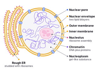
One of the key features of the nucleus is its double membrane, which is composed of two lipid bilayers. This double membrane acts as a barrier, separating the genetic material within the nucleus from the rest of the cell. This can be compared to a fortress wall, which protects the valuable contents within from external threats.
Inside the nucleus, the genetic material is organized into chromosomes, which can be thought of as the cell's instruction manual. This manual contains all the information needed to carry out the cell's functions and activities, and it is carefully regulated to ensure that the cell operates efficiently and effectively.
Inside the nucleus, there are a variety of other structures and organelles. These include the nucleolus, which is a non-membrane-bound organelle involved in the synthesis of ribosomes. The nucleolus is the site of rRNA synthesis and ribosome assembly, and it can be compared to a factory where parts are assembled and packaged before being sent out for use.
The nucleus also contains the nucleoplasm, which is the gel-like substance that fills the nucleus and surrounds the . The nucleoplasm can be compared to the fluid inside a water balloon, providing a medium for the many processes that occur within the nucleus.
The chromatin is visible when the cell is viewed under a microscope, and it contains the cell's genetic material. The chromatin can be compared to a tangled ball of yarn, with the genetic material wound tightly around proteins called histones. The chromatin helps to protect the DNA and regulate gene expression.
The nucleus also makes numerous small organelles called ribosomes, which are responsible for synthesizing proteins. Ribosomes can be found both in the cytoplasm and attached to the endoplasmic reticulum. They can be compared to workers in a factory, assembling the components needed to carry out the cell's functions.
Overall, the nucleus is an important organelle that plays a central role in the regulation of the cell's functions and the expression of its genetic information. The nucleolus and nucleoplasm, along with the chromatin and ribosomes, work together to ensure that the cell operates efficiently and effectively.
Whar Are Nuclear Pores?
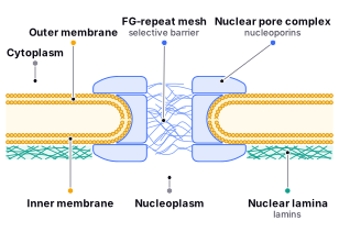
Nuclear pores are large, complex structures that facilitate the exchange of materials between the nucleus and the cytoplasm in eukaryotic cells. These pores are composed of multiple protein components and are embedded in the double membrane of the nuclear envelope.
The actual pore itself is formed by a large protein complex called the nuclear pore complex (NPC). The NPC is made up of over 30 different proteins, collectively called nucleoporins. These proteins form a mesh-like structure that spans the nuclear envelope, creating a passageway for molecules to move in and out of the nucleus.
Some nucleoporins are composed of multiple repeating units called FG-nucleoporin domains, which contain phenylalanine-glycine (FG) amino acid repeats. These domains are thought to create a selectively permeable barrier within the nuclear pore, allowing certain molecules to pass through while preventing others from doing so.
In addition to the NPC, the nuclear envelope itself also plays an important role in nuclear transport. The envelope is composed of two lipid bilayers, an inner and an outer membrane, which are connected by transmembrane proteins called nucleoporins. These proteins help to anchor the NPC to the nuclear envelope, as well as regulate the passage of molecules through the pore.
The nuclear lamina, a network of intermediate filaments that provides structural support to the nucleus, also plays a role in nuclear transport. The lamina is located on the inner side of the nuclear envelope and is composed of proteins called lamins. These proteins interact with the nucleoporins and other components of the nuclear envelope to help regulate the movement of molecules in and out of the nucleus.
Overall, nuclear pores are complex structures that allow for the regulated exchange of materials between the nucleus and the cytoplasm. The nucleoporins and other components of the nuclear envelope work together to create a selective barrier, while the nuclear lamina provides structural support and helps to regulate nuclear transport.
Find it in the figure
Coming soon for figure 3.4
Chromatin and Chromosomes
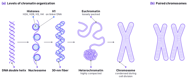
Chromatin is a complex of DNA and proteins found within the nucleus of eukaryotic cells, which makes up the chromosomes that carry the genetic information of the cell. Chromatin is composed of long strands of DNA wrapped around proteins called histones. The DNA strands in chromatin are tightly coiled and compacted, making it possible to fit a large amount of genetic information into a small space.
Chromosomes are the highly organized and compact structures that contain the genetic material of an organism. They are made up of chromatin and are visible under a microscope during cell division. Each chromosome is a single, continuous molecule of DNA, and each species has a characteristic number of chromosomes. For instance, humans have 46 chromosomes, while fruit flies have only eight.
In addition to histones, chromosomes also contain other proteins that help to organize and compact the chromatin. One of these proteins is called condensin, which plays a crucial role in the formation of chromosomes during cell division. Condensin helps to organize the chromatin into highly compacted and organized structures that can be easily segregated into daughter cells during cell division.
The structure of chromosomes can vary depending on the stage of the . During , the stage of the cell cycle when the cell is not actively dividing, chromosomes are in a more relaxed and extended state, allowing for gene expression and DNA replication. During cell division, however, the chromosomes become highly condensed and tightly packed, making it possible for them to be separated into daughter cells.
It is important to note that while chromosomes are highly organized and compact structures, they are not the only form of chromatin in the cell. Chromatin can exist in a range of different structures, from the loosely packed euchromatin to the highly compacted heterochromatin. These are two different types of chromatin that differ in their level of compaction and accessibility to enzymes and transcription factors. Euchromatin is loosely packed chromatin that is more accessible to these factors, allowing for the transcription of genes and the expression of specific traits or characteristics. Heterochromatin, on the other hand, is highly compacted and tightly packed chromatin that is less accessible to these factors, resulting in genes being less actively transcribed or silenced.
The balance between euchromatin and heterochromatin is crucial for the proper functioning of cells. During development and differentiation, specific genes need to be turned on or off to allow for the proper development of tissues and organs. This is accomplished through changes in chromatin structure, which can create a more open or closed chromatin state, depending on the gene's needs.
For example, during the development of the brain, specific genes involved in neuron development and function are turned on, while other genes that are not necessary for brain development are turned off. This process is regulated through changes in chromatin structure, which can create a more open chromatin state for the active genes and a more closed chromatin state for the inactive genes.
Understanding the organization and regulation of both chromatin and chromosomes is crucial for understanding the functioning of cells and the development of diseases such as cancer. Changes in the structure of chromatin and chromosomes can lead to alterations in gene expression, resulting in abnormal cell growth and function. Thus, research into the organization and regulation of chromatin and chromosomes is an important area of study in molecular biology.
Find it in the figure
Coming soon for figure 3.5
Histones
Histones are proteins that play a critical role in the structure and function of DNA in eukaryotic cells (cells that have a true nucleus and other membrane-bound organelles). They are responsible for compacting and organizing the DNA of the cell into a structure called chromatin.
Histones can be modified in various ways, including acetylation, methylation, and phosphorylation. These modifications can affect the structure of the chromatin and the accessibility of the DNA to the cell's machinery, such as enzymes that are involved in gene expression. For example, acetylation of histones is generally associated with more open, transcriptionally active chromatin, while methylation of histones is generally associated with more condensed, transcriptionally inactive chromatin.
Each type of histone plays a distinct role in regulating the structure and function of chromatin. For example, H2A and H2B histones are involved in stabilizing the nucleosome structure, while H3 and H4 histones play a crucial role in regulating gene expression.
H3 and H4 histones are subject to various modifications, such as acetylation, methylation, and phosphorylation, that can either enhance or inhibit gene expression. Acetylation of H3 and H4 histones, for instance, is associated with increased gene expression, while methylation can either increase or decrease gene expression depending on the specific location of the modification. Additionally, phosphorylation of H3 histones is involved in the regulation of cell division.
Another type of histone, H1, is not part of the nucleosome structure but is involved in organizing and compacting the chromatin into higher-order structures. H1 histones bind to the linker DNA between nucleosomes and play a role in the formation of higher-order chromatin structures, such as the 30-nm fiber.
Overall, the different types of histones play important and distinct roles in the regulation of chromatin structure and function. By modifying the histones and chromatin structure, cells are able to regulate gene expression and carry out a variety of functions necessary for cell growth, differentiation, and development.
The compact levels of chromosomes, chromatin, and histones are related to the organization of the cell's genetic material. Chromosomes are long strands of DNA that are found in the nucleus of a cell. They contain the cell's genetic information and are made up of chromatin, which is a complex of DNA and histones. The compact levels of chromosomes, chromatin, and histones are important for the cell's function because they allow the cell to fit a large amount of genetic material into a small space, such as the nucleus of a cell.
Overall, histones play a crucial role in the structure and function of DNA in eukaryotic cells. They help to compact and organize the DNA into a highly condensed structure called chromatin, which is essential for the cell's function and survival. Modifications to histones, such as acetylation and methylation, can affect the structure and accessibility of the DNA, which can have important consequences for gene expression and other cellular processes.
An analogy that may help to understand the idea of genetic material is to think of a recipe book. Just as a recipe book contains instructions for making a variety of dishes, the DNA in our cells contains the instructions for making all of the proteins and other molecules that are necessary for the cell to function. The recipes in a cookbook are written in a specific language and order, and the instructions in DNA are written in the language of nucleotide base pairs. Just as a cookbook can be passed down from one generation to the next, the genetic information in DNA is passed from one generation to the next through the process of reproduction. To protect the book (DNA), it's best not to bring it into the kitchen (Cytoplasm) where it could potentially be damaged (mutations). Instead, you quickly flip through the book, take a photo of the recipe you need (RNA), and return the book to a safe location (Nucleus). Then, you bring the copy of the recipe into the kitchen to use while cooking. This will ensure that the book stays in good condition while still allowing you to access the recipe.
Ribosomes
Ribosomes are small, but mighty structures found in all living cells. These tiny organelles are responsible for creating the proteins that make up much of the body's structure and perform many of its functions. Think of them like tiny protein factories that work tirelessly to produce the building blocks of life. Just as a carpenter needs wood, nails, and tools to build a house, a ribosome needs amino acids, messenger RNA, and transfer RNA to build a protein. Without ribosomes, the cell would not be able to create the proteins it needs to survive and carry out its functions. In this way, ribosomes are essential to all forms of life, from the tiniest bacterium to the largest mammal.
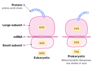
The structure of ribosomes varies depending on whether they are found in eukaryotic or prokaryotic cells. In eukaryotic cells have a ribosome size of 80S, ribosomes are composed of two subunits, a larger one called the 60S subunit and a smaller one called the 40S subunit. The 70S ribosome of prokartyoes is smaller and made up of a 50S and 30S subunits. The two subunits are held together by non-covalent bonds and can dissociate from each other when not in use.
Ribosomes are like chefs in a kitchen, reading a recipe book (mRNA) and using it to create a dish (protein). Just as a chef follows a recipe to add ingredients in a specific order, ribosomes read the mRNA to add amino acids in the correct sequence to build the protein. This process is called translation, and it is a vital part of gene expression. Ribosomes can synthesize a wide variety of proteins, from enzymes that catalyze chemical reactions to structural proteins that make up the cell's cytoskeleton. For example, ribosomes in pancreatic cells synthesize digestive enzymes that break down food in the digestive system, while ribosomes in muscle cells synthesize the proteins that make up muscle fibers. The process of protein synthesis is essential for the proper functioning of cells and is regulated by various factors, including hormones, nutrients, and environmental conditions.
Ribosomes are found in the cytoplasm of cells and are also found attached to the endoplasmic reticulum (ER). In eukaryotic cells, ribosomes on the surface of the ER are called the rough endoplasmic reticulum (RER) and are involved in the synthesis of proteins that are destined for the cell's membrane or for secretion. Ribosomes that are not bound to the ER are called free ribosomes and are involved in the synthesis of proteins that are to be used within the cell.
In summary, ribosomes are important organelles that play a central role in the synthesis of proteins. The structure and function of ribosomes varies depending on whether they are found in eukaryotic or prokaryotic cells.
Find it in the figure
Coming soon for figure 3.6
The “S” in Ribosolmal Weight
The “S” in ribosomal weight refers to the sedimentation coefficient of ribosomes, which is a measure of how quickly ribosomes sediment (or settle to the bottom of a tube) in a centrifuge. The sedimentation coefficient is often used as a measure of the size of ribosomes and other cellular particles.
Ribosomes can be isolated from cells and then purified by centrifugation. The purified ribosomes are then suspended in a solution and subjected to a process called ultracentrifugation. During ultracentrifugation, the sample is placed in a tube and spun at very high speeds in a centrifuge. The ribosomes sediment to the bottom of the tube based on their size and shape, with larger ribosomes sedimenting more quickly than smaller ones. The rate at which the ribosomes sediment can be measured using a device called a spectrophotometer, which measures the amount of light absorbed by the sample as it is spinning in the centrifuge. The sedimentation coefficient (S value) is then calculated based on the rate at which the ribosomes sediment and their size.
Prokaryotic ribosomes are smaller than eukaryotic ribosomes. This is because prokaryotes are generally smaller and less complex than eukaryotes, and their ribosomes reflect this difference in size. The ribosomes of mitochondria, which are the energy-producing organelles found in eukaryotic cells, are similar in size to prokaryotic ribosomes. This is thought to be due to the fact that mitochondria have a prokaryotic ancestry and have retained some characteristics of prokaryotes, including the size of their ribosomes.
The Mitochondria
To learn about the Mitochondria we, first of all, have to learn about the Endosymbiotic theory. The endosymbiotic theory is a scientific theory that explains the origin of mitochondria, which are small organelles found in the cells of most eukaryotes, including animals, plants, and fungi. According to this theory, mitochondria are the descendants of ancient bacteria that were engulfed by a larger host cell millions of years ago.
The story of the endosymbiotic theory begins billions of years ago, when the Earth was still a young planet. At this time, life on Earth consisted of simple, single-celled organisms known as prokaryotes. These organisms were small and relatively simple in structure, with no defined nucleus or other specialized organelles.
One day, a prokaryotic cell known as a host cell encountered a smaller prokaryotic cell known as a bacterium. The host cell was much larger than the bacterium, and it had the ability to engulf and incorporate other cells into its own structure. So, the host cell engulfed the bacterium, trapping it within its own membrane.
At first, the host cell saw the bacterium as a threat, and it attempted to destroy it using its own cellular machinery. However, the bacterium was able to resist these attempts and survive inside the host cell. Over time, the host cell began to recognize the bacterium as a beneficial symbiont, and it began to allow the bacterium to live and reproduce within its own membrane.
As the bacterium reproduced and passed its genetic material down to its offspring, it began to evolve and adapt to its new environment inside the host cell. It developed new functions and abilities that helped it to survive and thrive within the host cell. Eventually, the bacterium became so integrated into the host cell's functions that it became an essential part of the host cell's own structure and metabolism.
Today, the descendants of these ancient bacteria can be found in the cells of most eukaryotes, where they are known as mitochondria. They are responsible for producing energy for the cell through a process called cellular respiration, and they play a crucial role in the overall health and function of the cell.
So, the endosymbiotic theory is a fascinating story of how two simple, single-celled organisms came together in a symbiotic relationship that would ultimately shape the course of evolution on Earth. It is a testament to the incredible adaptability and resilience of life, and it serves as a reminder of the complex and interconnected nature of the living world. Now, let's talk about modern Mitochondria:
Mitochondria are specialized organelles found in both plant and animal cells. They are commonly known as the “powerhouses” or “energy factories” of the cell due to their role in producing the cell's main energy currency, adenosine triphosphate (ATP). Mitochondria are responsible for the process of cellular respiration, which involves converting nutrients, such as glucose, into ATP. This process uses oxygen and produces carbon dioxide as a waste product. The ATP produced is then used to fuel a wide range of cellular processes, including muscle contraction, cellular communication, and cellular maintenance. In addition to their role in energy production, mitochondria also play a role in other processes such as apoptosis (programmed cell death) and the synthesis of certain lipids and hormones.
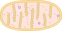
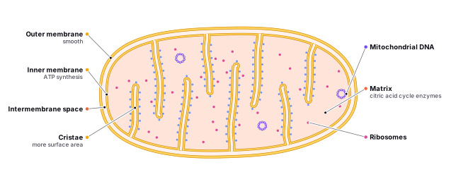
The structure of mitochondria is complex, and they are composed of a variety of proteins, lipids, and nucleic acids. Mitochondria are oval-shaped, double-membrane organelles with their own ribosomes and DNA. They have a double membrane, with the inner membrane being more highly folded than the outer membrane. The outer membrane is smooth, while the inner membrane is highly folded and contains numerous infoldings called cristae. The inner membrane of the mitochondria is where most of the ATP production occurs, with the cristae providing a large surface area for enzymes involved in the process. The area surrounded by the cristae is known as the mitochondrial matrix, which contains enzymes involved in the citric acid cycle and other biochemical pathways that produce ATP.
The two membranes of the mitochondria are composed of phospholipids and embedded with proteins. The outer membrane is porous and allows for the passage of small molecules, while the inner membrane is highly selective and contains transport proteins that regulate the passage of molecules, such as ions and metabolites, in and out of the matrix. The selective nature of the inner membrane allows for the establishment of an electrochemical gradient that drives the synthesis of ATP.
The inner membrane is responsible for several important functions in the mitochondria. It is the site of the electron transport chain, which is responsible for generating most of the cell's ATP (the main energy currency of the cell). The electron transport chain involves a series of proteins that transfer electrons from one molecule to another, generating a proton gradient that is used to produce ATP. This process requires a large surface area in order to accommodate all of the proteins that are involved in the electron transport chain.
In addition to the electron transport chain, the inner membrane also encloses the matrix, the site of the citric acid cycle, also known as the Krebs cycle. This is a series of reactions that takes place in the mitochondria, in which glucose and other nutrients are broken down to produce energy in the form of ATP.
The outer membrane of the mitochondria does not need to be folded because it does not have as many functions as the inner membrane. Its main role is to act as a barrier, separating the inside of the mitochondria from the rest of the cell. It does not need to have a large surface area for carrying out reactions, so it does not need to be folded.
Mitochondria are found in different numbers and concentrations in different cell types, depending on their energy requirements. For example, muscle cells have a high concentration of mitochondria to support the energy demands of muscle contraction, while other cells may have fewer mitochondria. Mitochondria are also able to divide and replicate themselves, allowing cells to increase their energy-producing capacity as needed.
Mitochondria are found in most eukaryotic cells, with the exception of red blood cells, which do not have mitochondria. They are particularly abundant in cells that have high energy demands, such as muscle cells and nerve cells.
In conclusion, the function of mitochondria in the cell is to produce ATP, the cell's main energy currency, through cellular respiration. The structure of mitochondria includes a double membrane with the inner membrane containing cristae that provide a large surface area for ATP production, transport proteins, and the mitochondrial matrix containing enzymes for the biochemical pathways that produce ATP. The number and concentration of mitochondria in different cell types reflect their energy requirements, with muscle cells having a higher concentration of mitochondria. Understanding the structure and function of mitochondria is crucial for understanding cellular energy metabolism and the development of diseases related to mitochondrial dysfunction.
Find it in the figure
Coming soon for figure 3.7
Peroxisomes
Peroxisomes are small, single-membrane-bound organelles found in the cytoplasm of eukaryotic cells. They are similar in size and shape to lysosomes and are also involved in the breakdown of biomolecules.
Peroxisomes are characterized by the presence of enzymes that use oxygen to break down certain biomolecules, such as fatty acids and amino acids. One of the main functions of peroxisomes is to detoxify harmful substances, such as alcohol, by converting them into more harmless compounds. They also play a role in the synthesis of certain lipids, such as plasmalogens, which are important for the proper functioning of cell membranes.
The structure of peroxisomes is relatively simple, and they are composed of a single membrane that encloses the enzymes and other molecules that are involved in their functions. Peroxisomes are formed from the endoplasmic reticulum (ER) and are similar in size and shape to lysosomes. However, unlike lysosomes, which are formed from the Golgi apparatus, peroxisomes are not involved in the degradation of large molecules such as proteins and polysaccharides.
Peroxisomes are found in most eukaryotic cells, and their functions vary depending on the type of cell and the specific needs of the organism. They are particularly important in the liver, where they play a role in the detoxification of harmful substances.
Animal vs. Plant Cells
Plant and animal cells are both eukaryotic cells, meaning they have a nucleus and membrane-bound organelles. However, there are several significant differences between the two cell types in terms of structure and function.
- Cell wall: One of the most noticeable differences between plant and animal cells is the presence of a rigid cell wall in plant cells. The cell wall is made primarily of cellulose, a complex carbohydrate, and provides structural support and protection for the plant cell. Animal cells, on the other hand, lack a cell wall and are surrounded only by a plasma membrane.
- Vacuoles: Both plant and animal cells contain vacuoles, which are membrane-bound sacs used for storage and waste disposal. However, plant cells generally have a large central vacuole that occupies a significant portion of the cell's volume. This central vacuole stores water, nutrients, and waste products and also helps maintain turgor pressure, which gives the plant cell its shape and rigidity. In contrast, animal cells have smaller vacuoles that are more dispersed throughout the cell.
- Plastids: Plant cells contain plastids, specialized organelles involved in the synthesis and storage of pigments, starch, and lipids. The most well-known plastid is the chloroplast, which contains the green pigment chlorophyll and is responsible for photosynthesis, the process by which plants convert sunlight into chemical energy. Animal cells do not have plastids, as they do not perform photosynthesis.
- Centrioles: Animal cells contain centrioles, which are cylindrical structures that play a crucial role in cell division by organizing the microtubules that form the spindle fibers. Plant cells do not have centrioles; instead, they rely on other structures to organize the spindle fibers during cell division.
- Lysosomes: Lysosomes are membrane-bound organelles that contain enzymes responsible for breaking down waste materials and cellular debris. While animal cells typically contain lysosomes, plant cells often rely on other organelles, such as vacuoles and peroxisomes, for similar functions.
- Cytoskeleton: Both plant and animal cells have a cytoskeleton, a network of protein fibers that provide structural support and play a role in cell movement and division. However, the cytoskeleton in plant cells is generally less extensive and more rigid than in animal cells, which reflects the different requirements for cell shape and movement.
- Shape and size: Plant cells are typically more rigid and rectangular or square in shape due to the presence of the cell wall, while animal cells are more flexible and can adopt a variety of shapes. Plant cells are generally larger than animal cells, with the central vacuole occupying a significant portion of the cell's volume.
In summary, plant and animal cells share many similarities as eukaryotic cells, but there are key differences in their structure and organelles, such as the presence of a cell wall, plastids, and a large central vacuole in plant cells, and centrioles and lysosomes in animal cells. These differences reflect the unique functions and requirements of each cell type.
The Chloroplast
Find it in the figure
Coming soon for figure 3.8
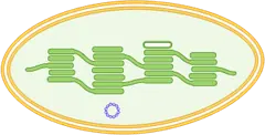
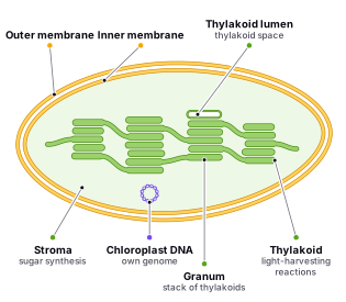
The chloroplast is a specialized organelle found in plant cells and some other types of organisms, such as algae. It is responsible for photosynthesis, the process by which plants, algae, and some bacteria convert light energy into chemical energy in the form of glucose. Chloroplasts contain pigments, such as chlorophyll, that absorb light energy and use it to convert water and carbon dioxide into glucose and oxygen through the light-dependent reactions and the Calvin cycle. Chloroplasts are found in the cells of plants and algae and are typically located in the mesophyll tissue of leaves, where they are exposed to light. They are shaped like discs and are surrounded by a double membrane. Inside the chloroplast, there are thylakoid membranes, where the light-dependent reactions of photosynthesis occur, and a stroma, where the light-independent reactions of photosynthesis take place.
The structure of a chloroplast can vary depending on the type of plant or algae it is found in, but all chloroplasts have a few key components:
- The outer membrane encloses the chloroplast and is made up of lipids.
- The inner membrane surrounds the stroma, which is the fluid-filled space inside the chloroplast. The inner membrane is also made up of lipids and contains proteins that are involved in the photosynthetic process.
- The thylakoid membrane is found inside the inner membrane and is made up of proteins and pigments such as chlorophyll. Thylakoids are arranged in stacks called grana.
- The thylakoid lumen is the space inside the thylakoid membrane and contains water.
- The stroma contains enzymes that are involved in the synthesis of glucose from carbon dioxide and water.
During photosynthesis, light energy is absorbed by pigments such as chlorophyll, which are located in the thylakoids. This energy is used to convert water and carbon dioxide into glucose and oxygen. The glucose is then used by the plant for energy, and the oxygen is released into the atmosphere as a byproduct of the reaction.
Section 3.4
The Endomembrane System
The endomembrane system is a group of interconnected structures within the cell that are involved in the synthesis, modification, and transport of various molecules. It includes the endoplasmic reticulum (ER), Golgi apparatus, lysosomes, vesicles, and the plasma membrane. These structures work together to create, modify, and transport proteins and lipids within the cell, as well as between cells.
The endoplasmic reticulum (ER) is a network of flattened membrane-bound sacs and tubes that is involved in the synthesis and modification of proteins and lipids. There are two types of ER: smooth endoplasmic reticulum (SER) and rough endoplasmic reticulum (RER).
The smooth endoplasmic reticulum (SER) lacks ribosomes and appears smooth under a microscope. It is involved in the synthesis and modification of lipids, including phospholipids and cholesterol, which are important components of cell membranes. The SER also plays a role in the detoxification of drugs and other harmful substances in liver cells.
On the other hand, the rough endoplasmic reticulum (RER) is involved in protein synthesis and modification. It is called “rough” because it is studded with ribosomes on its outer surface, giving it a bumpy appearance under a microscope. The ribosomes attached to the RER are the site of protein synthesis, and the proteins are then transported into the lumen of the RER, where they undergo further modification and folding.
The structural differences between the two types of ER are due to their different functions. The ribosomes attached to the RER are responsible for the synthesis of proteins, while the lack of ribosomes on the SER allows it to carry out its lipid synthesis and detoxification functions without interference.
An analogy for the difference between the two types of ER could be a factory with two different assembly lines. The RER would be the assembly line that produces products (proteins) with the help of specialized workers (ribosomes), while the SER would be the assembly line that produces the materials (lipids) needed for the products to be made, without the need for specialized workers.
Overall, the endoplasmic reticulum is a crucial part of the endomembrane system that plays a vital role in protein and lipid synthesis, modification, and transport within the cell.
The ER Signal Sequence
The ER signal sequence is a sequence of amino acids that is found in the N-terminus (beginning) of proteins that are destined for the endoplasmic reticulum (ER). This sequence serves as a signal for the protein to be transported into the ER by a process called protein targeting or protein sorting.
The main purpose of the ER signal sequence is to ensure that the protein is properly folded and modified within the ER before it is transported to its final destination, either within the cell or to the cell surface. This is important because many proteins require proper folding and modification to function correctly. For example, some proteins require the addition of carbohydrate groups to function properly, while others require disulfide bonds to stabilize their structure.
The ER signal sequence is typically composed of a stretch of hydrophobic amino acids, such as leucine, valine, and methionine. These hydrophobic amino acids interact with the lipid bilayer of the ER membrane and help the protein to be inserted into the membrane. The signal sequence also contains positively charged amino acids, such as lysine and arginine, which interact with the negatively charged phospholipids in the ER membrane.
Once the protein with the ER signal sequence is transported into the ER, the signal sequence is cleaved off by a signal peptidase enzyme. The remaining protein is then folded and modified in the ER, including the addition of carbohydrate groups and disulfide bonds, before it is transported to its final destination.
Defects in the ER signal sequence and protein folding process can lead to a variety of diseases, including cancer and neurodegenerative disorders. For example, misfolded proteins in the ER can trigger a response called the unfolded protein response (UPR), which can lead to cell death if not properly regulated.
In summary, the ER signal sequence plays a critical role in ensuring that proteins are properly folded and modified within the ER before they are transported to their final destination, and defects in this process can lead to a variety of diseases.
The Golgi Apparatus
The Golgi apparatus is a stack of flattened membrane-bound sacs that is involved in the modification, sorting, and packaging of proteins and lipids. It is a crucial part of the secretory pathway, which is responsible for the transport of proteins and lipids from the ER to their final destinations within the cell or to the cell surface.
The Golgi apparatus is made up of flattened stacks of membrane-bound sacs called cisternae. The cisternae are arranged in a linear fashion, with the cis face (entry face) at one end and the trans face (exit face) at the other end. The Golgi is therefore often referred to as having a “cis-to-trans” orientation. The cis face is where vesicles from the ER enter the Golgi, while the trans face is where vesicles carrying modified and packaged proteins and lipids exit the Golgi.
The Golgi is responsible for sorting, modifying, and packaging proteins and lipids that are produced in the endoplasmic reticulum (ER) and transported to the Golgi by vesicles. The Golgi apparatus performs a number of chemical modifications on these molecules, including:
- Glycosylation: the addition of carbohydrate groups to proteins and lipids. This is one of the most common modifications that occurs in the Golgi, and it is important for the proper folding and function of many proteins. For example, the ABO blood group antigens are determined by the addition of different carbohydrate groups to a protein called H antigen in the Golgi.
- Phosphorylation: the addition of phosphate groups to proteins and lipids. This modification can regulate protein activity and localization within the cell. For example, the addition of a phosphate group to a protein called tau in the Golgi can promote its binding to microtubules, which are important for maintaining the structure of nerve cells.
- Sulfation: the addition of sulfate groups to proteins and lipids. This modification can influence the ability of proteins to interact with other molecules within the cell. For example, sulfation of the carbohydrate groups on a protein called heparin in the Golgi is important for its anticoagulant activity.
- Proteolysis: the cleavage of proteins into smaller peptides or amino acids. This modification can regulate protein activity and function. For example, the processing of the precursor protein proinsulin in the Golgi produces the mature hormone insulin, which regulates glucose metabolism in the body.
Once the proteins and lipids have been modified in the Golgi, they are packaged into vesicles and transported to their final destination, either within the cell or to the cell surface. For example, proteins that are destined for secretion from the cell are packaged into vesicles that fuse with the plasma membrane and release their contents outside the cell.
In summary, the Golgi apparatus is a complex organelle that performs a variety of chemical modifications on proteins and lipids, including glycosylation, phosphorylation, sulfation, and proteolysis. These modifications are essential for the proper folding, function, and localization of these molecules within the cell. The Golgi also plays a critical role in sorting and packaging these molecules into vesicles for transport to their final destination.
Lysosomes and Vesicles
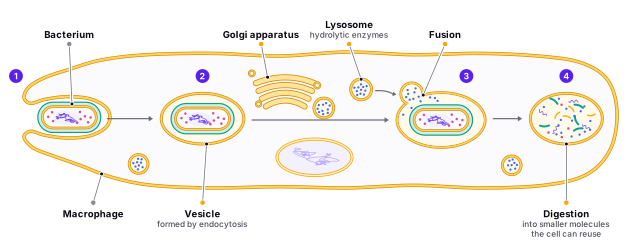
Lysosomes are small, membrane-bound organelles that contain hydrolytic enzymes that can break down a variety of biomolecules, including proteins, carbohydrates, and lipids. They are involved in the degradation of material taken up by the cell through endocytosis, as well as in the degradation of aged or damaged cell organelles.
The structure of lysosomes is unique in that they are formed by the fusion of vesicles produced by the Golgi apparatus with endosomes containing material that has been internalized by the cell. Once the lysosome is formed, it contains hydrolytic enzymes that can break down the material inside into smaller molecules that can be reused by the cell.
Lysosomes are essential for maintaining cellular homeostasis, as they are responsible for breaking down and recycling cellular waste products. Defects in lysosomal function can lead to a variety of lysosomal storage disorders, such as Tay-Sachs disease and Gaucher disease, where the accumulation of undegraded biomolecules in lysosomes can cause cellular damage and organ dysfunction.
Vesicles, on the other hand, are small, membrane-bound structures that transport materials within the cell. They can be formed by the Golgi apparatus, the RER, or by the plasma membrane through processes such as endocytosis or exocytosis. Vesicles can transport materials to other parts of the cell, or they can fuse with the plasma membrane to release their contents outside of the cell.
The structure of vesicles can vary depending on their function. For example, synaptic vesicles in nerve cells are specialized vesicles that store neurotransmitters, while secretory vesicles in glandular cells store and secrete hormones.
Vesicles are medically relevant because they are involved in a variety of cellular processes that can impact human health. For example, defects in vesicle transport can lead to neurodegenerative disorders such as Parkinson's and Alzheimer's disease. Additionally, viral infections can hijack the vesicle transport system to facilitate their own replication and spread within the body.
An analogy for lysosomes and vesicles could be a garbage disposal and a mail carrier, respectively. Lysosomes act like a garbage disposal, breaking down and recycling waste products within the cell. Vesicles act like a mail carrier, transporting materials to different parts of the cell or outside of the cell, like a mail carrier delivering packages to different addresses.
Find it in the figure
Coming soon for figure 3.9
Section 3.5
Cytoskeleton
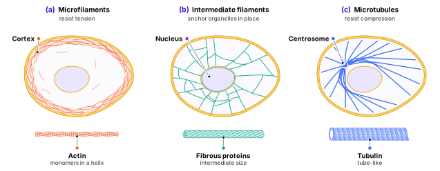
The cytoskeleton is a network of protein filaments within the cell that provides structural support, helps to maintain cell shape, and plays a role in cell division and movement. There are three main types of protein filaments that make up the cytoskeleton: microtubules, microfilaments, and intermediate filaments.
Microtubules are long, thin, tube-like structures made up of protofilaments of the protein tubulin. They are found throughout the cell, but are particularly abundant near the nucleus and in the periphery of the cell. Microtubules are involved in the maintenance of cell shape, the movement of organelles within the cell, and the separation of chromosomes during cell division. Microtubules are also involved in the formation of the mitotic spindle, which is essential for the proper segregation of chromosomes during cell division.
Microtubules have a variety of specific uses within the cell. For example, they form the axonemes of cilia and flagella, which are involved in the movement of cells and the movement of fluids over the surfaces of cells. They also provide tracks for the transport of materials within the cell, including the transport of neurotransmitter-containing vesicles in nerve cells.
Microfilaments are thin, rope-like structures made up of the protein actin. They are found throughout the cell, but are particularly abundant near the plasma membrane. Microfilaments are involved in cell movement and in the contraction of muscle cells. Microfilaments are also involved in cell division and in the formation of the contractile ring during , which helps to divide the cell into two daughter cells.
Microfilaments have a variety of specific uses within the cell. For example, they form the contractile ring in the cleavage furrow during cell division, which helps to separate the cell into two daughter cells. They also form the structural framework for microvilli, which are finger-like projections on the surface of cells that increase the surface area for absorption.
Intermediate filaments are thicker than microfilaments, thinner than microtubules, and more stable than both. They are made up of a variety of proteins and are found throughout the cell, but are particularly abundant in cells that are subjected to mechanical stress, such as epithelial cells and muscle cells. Intermediate filaments are involved in the maintenance of cell shape, as well as in the attachment of organelles to the cytoskeleton.
Intermediate filaments have a variety of specific uses within the cell. For example, they provide the structural support for the nuclear envelope, which surrounds and protects the genetic material of the cell. They also provide the structural support for desmosomes, which are specialized cell junctions that help to anchor cells together in tissues that are subjected to mechanical stress, such as the skin and heart tissue.
In summary, the cytoskeleton is a complex network of protein filaments that provides structural support, maintains cell shape, and plays a critical role in cell division and movement. The three main types of protein filaments in the cytoskeleton - microtubules, microfilaments, and intermediate filaments - differ in their structure, function, and location within the cell, and each has specific uses within the cell.
Find it in the figure
Coming soon for figure 3.10
Microtubules
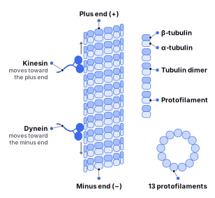
Microtubules are long, thin, tube-like structures that are an important component of the cytoskeleton in eukaryotic cells. They are composed of protein subunits called tubulin, which polymerize to form the microtubule.
Microtubules have a number of important functions in the cell, including:
- Maintaining the shape and structure of the cell: Microtubules help to maintain the overall shape of the cell and provide structural support for organelles and other structures within the cell. For example, microtubules help to maintain the shape of neurons, which have long extensions called axons and dendrites that require a stable framework to support them.
- Participating in cell division: During cell division, microtubules help to separate the duplicated chromosomes and distribute them to the two daughter cells. They do this by forming the mitotic spindle, a structure that helps to pull the chromosomes apart. Microtubules also play a role in cytokinesis, the process of dividing the cell into two daughter cells.
- Transporting materials within the cell: Microtubules can also act as tracks for motor proteins, which transport vesicles, organelles, and other materials within the cell. For example, kinesin and dynein are motor proteins that move along microtubules and transport vesicles containing neurotransmitters in nerve cells.
- Regulating gene expression: Microtubules can also play a role in regulating gene expression by helping to position the chromosomes within the cell. This positioning can affect how genes are expressed and can influence cell fate and differentiation.
In addition to these functions, microtubules are involved in a variety of cellular processes, such as the maintenance of cell polarity, the formation of cilia and flagella, and the movement of cells. Defects in microtubule function can lead to a variety of diseases, including cancer, neurodegenerative disorders, and ciliopathies, which are disorders caused by defects in cilia function. An analogy for microtubules could be a scaffold or support beam in a building. Just like a scaffold provides support and maintains the structure of a building, microtubules provide support and maintain the structure of the cell.
Another way to understand the structure and function of microtubules, is you can think of them as the “highways” of the cell. Just as highways help to transport people and goods from one place to another, microtubules help to transport materials within the cell. They also help to maintain the overall structure of the cell and play a role in important processes such as cell division.
There are several different types of motor proteins that can move along microtubules, including kinesin and dynein. Kinesin moves towards the plus end of the microtubule, while dynein moves towards the minus end. Motor proteins are important for a variety of processes, including the transport of vesicles and organelles within the cell, and the movement of cilia and flagella.
Find it in the figure
Coming soon for figure 3.11
Motor Proteins
Kinesin and dynein are two types of motor proteins that are responsible for transporting cargo within cells along microtubules. Kinesin moves towards the plus end of the microtubule, while dynein moves towards the minus end, allowing them to move cargo in opposite directions.
An analogy for kinesin and dynein could be like trains on a railway system. Just like trains travel in opposite directions on a railway system, kinesin and dynein move in opposite directions along microtubules to transport cargo within the cell.
Kinesin is composed of two globular heads and a long, flexible stalk that connects them. The heads of kinesin bind to the microtubule and use energy from ATP hydrolysis to “walk” towards the plus end of the microtubule, pulling cargo along with them. An example of kinesin in action is the transport of synaptic vesicles containing neurotransmitters in nerve cells. Kinesin moves these vesicles from the cell body to the axon terminal, where they are released to transmit signals to other neurons or to muscles.
Dynein is similar to kinesin in that it is composed of two globular heads and a long, flexible stalk, but it moves in the opposite direction towards the minus end of the microtubule. Dynein is responsible for moving cargo such as vesicles and organelles towards the center of the cell or towards the (MTOC). An example of dynein in action is the movement of chromosomes during cell division. Dynein helps to position the mitotic spindle and move the chromosomes towards the MTOC for proper segregation.
Kinesin and dynein are also involved in the movement of cilia and flagella, which are hair-like structures that extend from the surface of cells and are involved in movement and sensing. Defects in kinesin, dynein, or microtubules can lead to problems with ciliary or flagellar movement, resulting in diseases known as ciliopathies, which can affect various organs and tissues in the body. For example, defects in the cilia of the respiratory tract can lead to chronic respiratory infections, while defects in the cilia of the kidneys can cause polycystic kidney disease.
In addition, defects in the function of kinesin, dynein, or microtubules can also contribute to neurodegenerative disorders such as Alzheimer's disease and Parkinson's disease, which involve the abnormal accumulation of proteins in the brain. Understanding the functions of kinesin, dynein, and microtubules is therefore crucial for understanding the underlying mechanisms of these diseases and developing potential treatments.
Cilia and Flagella
Cilia and flagella are fascinating structures with important functions in movement, sensing, and signaling in eukaryotic cells. To better understand these structures, it helps to break down their individual components and functions.
Cilia are hair-like structures that protrude from the surface of cells and are involved in various physiological processes. They consist of microtubules arranged in a 9 + 2 pattern, moved by proteins called dyneins. Kinesin and dynein motor proteins move along the microtubules, with kinesin moving towards the plus end and dynein moving towards the minus end. The movement of dynein results in the cilium bending and moving in a waving motion. Cilia can be found in many different tissues and organs, including the respiratory and reproductive tracts and the inner ear.
Flagella, on the other hand, are whip-like structures that are longer than cilia and used for propulsion. They are also composed of microtubules arranged in a 9 + 2 pattern, moved by dyneins. Flagella are generally found only in sperm cells, where they help the sperm swim towards the egg.
The bacterial flagellum is similar in function to eukaryotic cilia and flagella, but it is composed of a single, long filament of flagellin anchored to the cell membrane. The flagellum is powered by a motor protein called the flagellar motor, which uses the energy from a proton gradient to rotate the filament.
Primary cilia, on the other hand, are small, hair-like structures that are found on the surface of many different types of cells in the body. They are similar in structure to other cilia, but they are generally shorter and have a different function. Primary cilia consist of microtubules arranged in a 9 + 0 pattern and are involved in sensory functions, signal transduction, and development. They are found on many different types of cells, including epithelial cells, nerve cells, and muscle cells.
It is important to note that defects in cilia and flagella function can lead to a variety of medical conditions, including ciliopathies, neurodegenerative disorders, and male infertility. Therefore, understanding the structure and function of these fascinating structures can have important medical implications.
Cilia are found in a number of different tissues and organs, including:
- The respiratory tract: Cilia lining the respiratory tract help to move mucus and trapped particles out of the lungs.
- The reproductive tract: Cilia in the female reproductive tract help to move eggs and sperm towards the uterus and Fallopian tubes.
- The inner ear: Cilia in the inner ear help to detect sound and maintain balance.
Flagella on the other hand can mainly be found in the tail of the sperm, it helps it propel towards the egg.
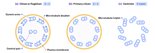
Find it in the figure
Coming soon for figure 3.12
The Microtubule-Organizing Center
The microtubule-organizing center (MTOC) is a structure found in eukaryotic cells that is responsible for organizing and maintaining the microtubules in the cell. The MTOC is typically located near the center of the cell and is composed of a group of proteins called the centrosome, which is made up of two centrioles.
Each centriole is made up of nine triplets of microtubules arranged in a circular fashion. The triplets are connected by accessory proteins, which help to hold the microtubules in place. The centrioles are surrounded by a layer of protein called the pericentriolar material (PCM), which is involved in the nucleation of microtubules.
The centrosome is the primary site for the nucleation of microtubules in the cell, and the centrioles are responsible for the synthesis of the microtubules. The centrosomes replicate during the cell cycle and produce daughter centrioles, which are used to form the mitotic spindle during cell division. The mitotic spindle is a structure made up of microtubules that helps to separate the duplicated chromosomes and distribute them to the daughter cells.
Defects in the function of the MTOC can result in a number of diseases, including ciliopathies, which are caused by defects in the structure or function of cilia and flagella, as well as neurodegenerative disorders. Additionally, the MTOC is a target for cancer therapies, as the mitotic spindle is essential for cell division and proliferation, and disrupting its function can lead to cell death.
Microfilaments
Microfilaments are thin, rod-like structures that are an important component of the cytoskeleton in eukaryotic cells. They are composed of protein subunits called actin, which polymerize to form the microfilament.
Microfilaments have a number of important functions in the cell, including:
- Maintaining the shape and structure of the cell: Microfilaments, also known as actin filaments, are one of the main components of the cytoskeleton, which is a network of protein filaments within the cell that provides structural support and helps to maintain cell shape. Microfilaments are responsible for maintaining the overall shape of the cell and for providing structural support for organelles and other structures within the cell. For example, microfilaments help to maintain the shape of red blood cells, which are able to squeeze through narrow capillaries due to their flexible shape.
- Microfilaments are also important for the movement of cells. They are involved in processes such as cell division, muscle contraction, and the movement of organelles within the cell. During cell division, microfilaments help to form the contractile ring, which helps to divide the cell into two daughter cells. In muscle cells, microfilaments work together with myosin motor proteins to generate the force needed for muscle contraction. Microfilaments also help to transport organelles within the cell, such as vesicles and mitochondria
- Participating in signaling pathways: Microfilaments can also play a role in signaling pathways, which are pathways that transmit information within the cell. For example, microfilaments can interact with other proteins to regulate the activity of enzymes involved in signaling pathways. Microfilaments can also be involved in the formation of membrane protrusions, such as filopodia and lamellipodia, which are important for cell movement and for interactions between cells.
To understand the structure and function of microfilaments, you can think of them as the “scaffolding” of the cell. Just as scaffolding helps to support the structure of a building, microfilaments help to support the structure of the cell and play a role in important processes such as cell movement.
Actin and microfilaments are important components of the cytoskeleton, a network of fibers that helps to maintain the shape and structure of eukaryotic cells. They are made up of protein filaments that are arranged in a linear fashion and are essential for many different functions in the cell.
Actin is a protein that is found in all eukaryotic cells and is an important component of the cytoskeleton. It is composed of a chain of monomeric subunits called actin monomers, which are arranged in a helical fashion. There are two main types of actin found in cells: filamentous actin (F-actin) and globular actin (G-actin).
F-actin is the filamentous form of actin and is made up of long chains of actin monomers. It is an important component of the cytoskeleton and is involved in many different functions in the cell, including:
- Cell shape: F-actin, or filamentous actin, is a major component of the cytoskeleton and is responsible for maintaining the shape of the cell. It forms a dynamic scaffold that provides support and structure to the cell, allowing other cell components to attach to it. F-actin also helps to maintain the integrity of the plasma membrane by anchoring it to the cytoskeleton.
- Movement: F-actin plays a critical role in the movement of cells and organelles within the cell. It is involved in the formation of contractile structures, such as the contractile ring during cytokinesis and the actin-myosin complex in muscle cells. In addition, F-actin is involved in the movement of organelles and vesicles within the cell, through interactions with motor proteins such as myosin.
- Signal transduction: F-actin is involved in signaling pathways that control various cellular processes. For example, F-actin is involved in the activation of Rho GTPases, which are signaling molecules that regulate actin dynamics and are involved in processes such as cell division and migration. F-actin is also involved in the activation of transcription factors, which are proteins that control the expression of genes in response to specific signals. In addition, F-actin is involved in the formation of specialized structures within the cell, such as filopodia and lamellipodia, which are involved in processes such as cell migration and adhesion.
G-actin is a small, globular protein that is made up of individual actin monomers. It is typically found in the cytosol of eukaryotic cells, where it can exist in equilibrium with F-actin, the filamentous form of actin.
G-actin plays a crucial role in the formation of F-actin filaments, as it is the building block for the filaments. Actin nucleation factors, such as Arp2/3 and formins, bind to G-actin and promote the formation of F-actin filaments by polymerizing the individual actin monomers into long chains.
In addition to its role in filament formation, G-actin also plays a role in various cellular processes, such as cell migration, cell division, and intracellular transport. For example, G-actin is involved in the formation of pseudopodia, which are temporary protrusions that allow cells to move in a particular direction. G-actin is also involved in the formation of contractile rings during cytokinesis, the process by which a cell divides into two daughter cells.
The concentration of G-actin in the cytosol is tightly regulated, as it is important for maintaining the balance between G-actin and F-actin. Alterations in the balance can lead to changes in the structure and function of the cell. For example, changes in the concentration of G-actin have been associated with various diseases, including cancer and heart disease.
Overall, G-actin is a crucial protein in the cytosol of eukaryotic cells, where it serves as a building block for F-actin filaments and plays a role in various cellular processes. Its location and function are tightly regulated, and alterations in its concentration can have significant consequences for the cell.
Intermediate filaments
Intermediate filaments are a diverse group of fibrous proteins that are an integral part of the cytoskeleton in eukaryotic cells. They are so named because of their intermediate size between microtubules and microfilaments. Intermediate filaments are found in the cells of animals, where they play a vital role in maintaining cellular structure and integrity. Unlike microtubules and microfilaments, intermediate filaments are not involved in cell division or movement but provide structural support to cells and tissues subjected to mechanical stress. Intermediate filaments are made up of a family of proteins that differ in their composition and structure depending on the cell type and tissue. They are organized into bundles, networks, and sheets, where they act as anchors to hold cells and tissues together. Intermediate filaments are present in a wide variety of cell types, including epithelial cells, muscle cells, and nerve cells. Their diverse structural properties allow them to fulfill a variety of roles in different cell types and tissues. Understanding the structure and function of intermediate filaments is crucial to understanding the behavior of cells and tissues under various physiological and pathological conditions.
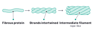
Find it in the figure
Coming soon for figure 3.13
Intermediate filaments have a number of important functions in the cell, including:
- Maintaining the shape and structure of the cell: Intermediate filaments provide mechanical support to cells and help to maintain their overall shape and structure. They form a network of filaments that is resistant to tensile forces, which can help cells to resist deformation and maintain their shape. Different types of intermediate filaments are found in different cell types, with some being particularly important for maintaining the shape of cells that are subjected to mechanical stress, such as skin cells or muscle cells.
- Participating in cell movement: While intermediate filaments are not as directly involved in cell movement as microtubules or microfilaments, they can still play a role in this process. For example, intermediate filaments have been shown to be important for the migration of some types of cells, such as fibroblasts. In addition, they are thought to be involved in the maintenance of the shape and structure of certain specialized structures involved in cell movement, such as the lamellipodia and filopodia that extend from the leading edge of migrating cells.
- Protecting the cell from mechanical stress: Intermediate filaments can help to protect cells from mechanical stress by providing a tough, flexible network that can withstand forces without breaking. This is particularly important in cells that are subjected to constant mechanical stress, such as skin cells or muscle cells. For example, intermediate filaments in muscle cells help to maintain the integrity of the sarcomere structure during muscle contraction, preventing damage to the muscle fibers.
To understand the structure and function of intermediate filaments, you can think of them as the “reinforcing bars” of the cell. Just as reinforcing bars help to strengthen a structure and protect it from stress, intermediate filaments help to maintain the overall shape of the cell and protect it from mechanical stress.
Section 3.6
Comparing Animal, Plant and Bacteria Cells
Animal cells:
- Lack a cell wall
- Have smaller cell size compared to plant cells
- Have centrioles, which are involved in cell division
- Have lysosomes, which contain enzymes that break down waste materials and cell debris
- Have mitochondria, which are responsible for generating energy in the form of ATP
- Do not have chloroplasts, which are responsible for photosynthesis in plant cells
Plant cells:
- Have a cell wall made of cellulose, which provides support and protection
- Have larger cell size compared to animal cells
- Do not have centrioles
- Have a central vacuole, which stores water and other substances
- Have chloroplasts, which are responsible for photosynthesis
- Do not have lysosomes
Bacteria cells:
- Have a cell wall made of peptidoglycan, which provides support and protection
- Can have various shapes, including spherical, rod-shaped, and spiral-shaped
- Do not have a defined nucleus; instead, their genetic material is found in a region called the nucleoid
- Do not have organelles like mitochondria or lysosomes
- Can have flagella, which are used for movement
- Can have pili, which are used for attachment and exchange of genetic material